
Howard Fischer
Uppsala, Sweden
Scribe: noun. A person who copies documents, especially a person who made handwritten copies before the invention of printing.
— Dictionary.com
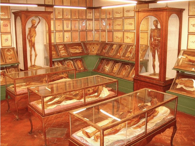
The first reliable anatomic drawings based on human dissections may have been those of Leonardo Da Vinci (1452–1519). Later, Andreas Vesalius (1514–1564), born in Brussels as Andries van Wesel and having taken a medical degree from the University of Pavia, produced a complete, seven-volume atlas of human anatomy based on his many dissections. This milestone was his On the Structure of the Human Body (1543). At about the same time, finely and accurately detailed wax models of the human body with thoracic, abdominal, and pelvic (male and female) organs in place were produced in Italy for medical student education.
Much later, photography and then roentgenography were adapted for medical education. An enormous breakthrough in diagnostic imaging—and secondarily for medical education— came in 1972 with the development of a practical computer-assisted tomography (CAT) machine. This technology enabled physicians to visualize an organ as a series of sections or slices (tomos—slice, section in Ancient Greek). The first CAT scanner arrived in the US in 1973. The 1979 Nobel Prize in Physiology or Medicine was awarded to its developers, physicist Allan Cormack and electrical engineer Godfrey Hounsfield.
With this brief background in mind, we turn to the teaching of anatomy at the medical school I attended in Belgium, where Vesalius himself once taught. Our anatomy building was called the “Vesalius Institute.” Our first human anatomy course involved learning osteology (bones) and myology (muscles). We studied most of the bones in a hands-on manner, with articulated and disarticulated skeletons. We were offered human skulls at a low cost for home study. The professor advised us to study any one of several standard anatomy texts (in French, in English and Latin, or in German). This was, fifty years ago, the traditional way to learn these subjects.
Our next anatomy course was the study of splanchnology (from the Latin splanchno meaning viscera). A different professor entered the auditorium—we were about 300 students—carrying a box with chalk in eight different colors. He drew on the blackboard drawings representing anatomical “cuts,” (anticipating CAT scan imaging) of the human body at various levels. The students had to copy these drawings using the exact same colors, proportions, and labels that the professor used. Interesting and instructive? Yes. However, the student was required to memorize these hundreds of drawings. I felt like a copy machine in this course. I thought about Saint Veronica,1 who wiped the face of the fallen Jesus on his way to Golgotha, and the image of Christ remained on the handkerchief that this good woman used. In our course, no textbook was recommended. These drawings, plus attendance at several autopsies, were the course.
The final examination consisted of reproducing, from memory, a number of these drawings. The specific drawings required were listed on index cards that each student picked, blindly, on entering the examination hall. For me, this course was an ordeal. I still have nightmares about it, and so do a few of my classmates.
Reference
- Catholic Saints Info Blog Archive. “Saint Veronica.” https://catholicsaints.info/saint-veronica/.
HOWARD FISCHER, MD, was a professor of pediatrics at Wayne State University School of Medicine, Detroit, Michigan. He has a longtime interest in things Belgian.