Howard Fischer
Uppsala, Sweden

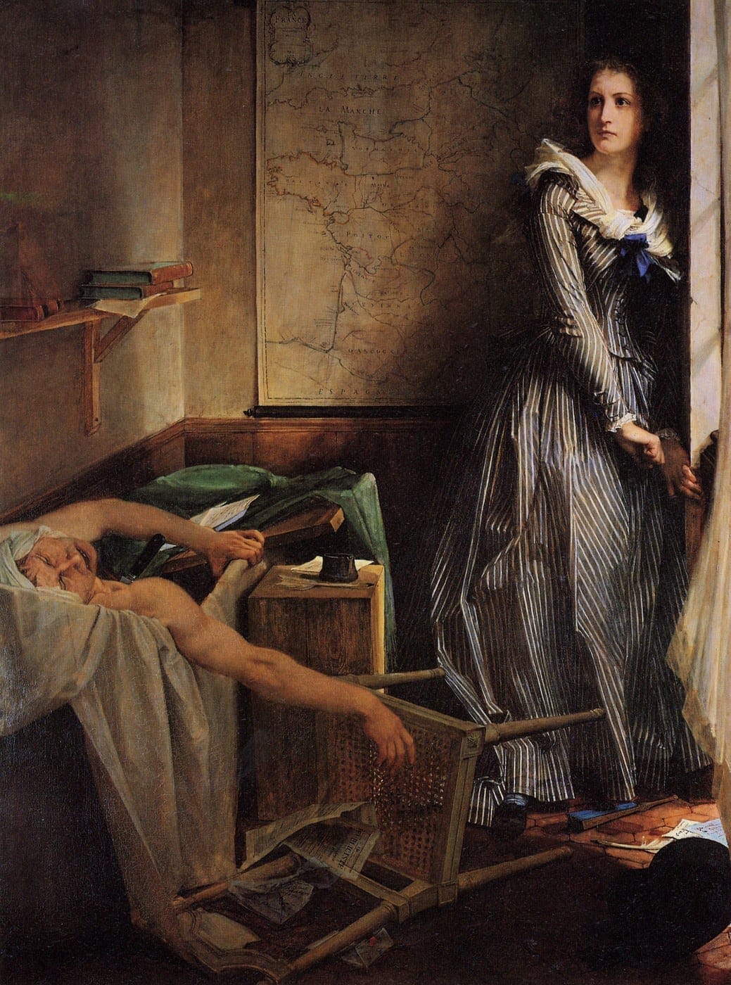
Jean-Paul Marat (1743-1793) was a practicing physician, scientist, and a leader of the French Revolution. He also suffered from a chronic, intractable skin condition, which troubled the last five years of his life. A tormenting itch caused him to spend whole days1 in his custom-made bathtub, from which he wrote revolutionary articles and received visitors. It was in this bathtub that he was murdered by Charlotte Corday (1768-1793), a scene immortalized in the painting Marat Assassiné by Jacques-Louis David.
Marat was around forty-five when the itching and vesico-bullous lesions began, first on the scrotum, then the groin and perineum, and later became widespread.2 His only relief came from spending long periods in his bath. Marat believed that his skin disease was caused by his earlier years as a fugitive hiding in the cellars and sewers of Paris.
There have been centuries of speculation about the nature of his disease. During his lifetime, Marat’s enemies claimed that his skin eruption was syphilitic, though none of the features of his rash (itch, duration) support this notion. Murphy3 states that Marat’s contemporaries believed he had leprosy, “and some people began to treat him as a leper,” but this too is an incorrect diagnosis. Scabies has been suggested as a cause, but scabies does not usually involve the face in adults, and, as a physician, Marat would have recognized it and used “the reliable form of therapy” that was then available.4 Marat’s own physicians diagnosed him with scrofula,5 but this condition of tuberculous lymph node involvement does not fit the clinical picture. Cohen and Cohen6 have made a case for seborrheic dermatitis, but the severity of Marat’s symptoms and description of his lesions make this questionable. Dermatitis herpetiformis, a chronic blistering condition, has also been suggested, but while pruritic, it does not involve the scrotum and is associated with dietary gluten intolerance (celiac disease).
Marat has been described as having an “insatiable thirst”14,15 that he could barely satisfy,16 a condition that seems to have been contemporaneous with his skin manifestations. He consumed large quantities of water mixed with almond paste, into which were ground bits of clay.17 Diabetes could account for Marat’s enormous thirst, but a condition such as diabetic candidiasis does not cause “ceaseless, generalized pruritus.”7 More importantly, it is unlikely that he would have survived five years of untreated, severe diabetes. His long survival with these symptoms also helps to exclude the diagnosis of pemphigus vulgaris.8 Hebra’s prurigo,9 another severely pruritic condition, begins in infancy and is papular, not vesicular in appearance. A psychogenic etiology for Marat’s itching has also been postulated.10
Eczema is the diagnosis favored by Cabanès,11 although most eczema first appears in childhood. Severe eczema, especially if complicated by the nodules of prurigo nodularis, may produce an incessant, intolerable itch that may be temporarily reduced by bathing. Wahlberg12 has proposed Sézary syndrome, a late form of mycosis fungoides or T-cell lymphoma, which affects the skin. He points out that the erythroderma (“red skin”) that is part of Sézary syndrome is not seen in David’s painting, though it is unknown whether the artist’s goal was a symbolic memorial on canvas or a faithful portrayal of dermatologic detail.
More recently, genomic analysis of a bloodstain left on papers at Marat’s assassination13 showed evidence of Malassezia restricta, a fungus implicated in seborrheic dermatitis, as well as Cutibacterium (formerly Proprionibacterium) acnes. There was no genetic evidence of syphilis, leprosy, tuberculosis, candida, or scabies. From this, the authors offered a possible diagnosis of seborrheic dermatitis with bacterial superinfection. However, seborrheic dermatitis is not as itchy as Marat’s condition, secondary infection is not common, and the distribution of lesions is not consistent with seborrheic dermatitis.
Might Marat’s excessive thirst point to yet another, more systemic diagnosis? Diabetes insipidus is a condition where antidiuretic hormone (ADH), which is produced in the hypothalamus and released into the bloodstream from the posterior part of the pituitary gland, is reduced, affecting a feedback loop that results in the kidneys producing large quantities of dilute urine and resulting thirst. The physiological mechanisms involved in this condition may point to a unifying diagnosis for Marat’s symptoms.
In 1937, Turkish dermatologist Hulusi Behçet published a paper19 describing patients who had oral and genital ulcers (“aphthae”) and eye inflammation. Ten years later the name “morbus Behçet” was proposed and accepted as the name of this group of findings.20 Behçet’s disease is a relapsing inflammatory syndrome of unknown origin that may involve multiple organ systems. The highest prevalence is seen in Japan, southeast Asia, the Middle East, and southern Europe—the path of the Silk Road. Its worldwide prevalence is 1:16000 to 1:20000 individuals with a slight male predominance. It typically appears in young adulthood, but may have its onset in childhood or in middle age.21 It is generally agreed that oral ulcers are a near-invariable finding. Ulcers also appear on the genitals, especially the scrotum or vulvae, in 80% of patients.22 There is eye inflammation in 60-70% of patients23 but eye involvement may not develop until ten years after the onset of other symptoms.24 The skin develops papulopustular lesions (60-80%) and the joints may be affected. Because blood vessels also become inflamed, the cardiovascular, renal, and gastrointestinal systems are also potential targets.
Up to 33% of patients have nervous system involvement,25 manifesting variably as headache, insomnia, cranial nerve palsies, cerebral venous sinus thrombosis, ataxia, and dementia.26 There have been a few reports of patients with Behçet’s disease involving the blood vessels supplying the pituitary gland, including a recent report of a forty-year-old man diagnosed with Behçet’s disease five years earlier and presenting with thirst, increased fluid intake, and frequent urination.27 He was diagnosed with diabetes insipidus induced by vasculitis of the blood vessels supplying the pituitary gland, which was confirmed by magnetic resonance imaging.
Marat was of Sardinian (i.e. southern European) heritage and a middle-aged man when his skin problems began. His headaches, insomnia, personality changes, and excessive thirst might all have been caused by neurologic manifestations of Behçet’s disease. His scrotal and perineal ulcers are compatible with the diagnosis of Behçet’s, as is his more diffuse skin condition. Arm and leg pains28,29 could have been manifestations of associated arthritis or of vasculitis compromising blood supply to limb muscles. Shearing30 states that Marat had “piercing yellow-gray eyes, infected with bile and blood,” which could indicate the anterior uveitis, common in Behçet’s disease. Marat was never described as having oral lesions, which prevents this diagnosis with great assurance, although his preference for liquids over solid food and his consumption of almond paste-clay water may have been a method of calming painful oral lesions.
The multisystemic nature of Behçet’s disease makes it a reasonable candidate to account for Marat’s physical and behavioral problems. Marat was indeed a man with many problems, as well as many virtues. Perhaps more astute medical detectives will join in the search for a diagnosis to fit this centuries-old question.
References
- Augustin Cabanès, “Quelle Était la Maladie de Marat?” J Maladies Cutanées et Syphilitiques, 4 (1894): 125-128.
- H.L. Cohen and E.L. Cohen, “Doctor Marat and his Skin,” Med Hist, 24 (1958): 281-286.
- Lisa C. Murphy, “The Itches of Jean-Paul Marat,”J Am Acad Derm 21, no. 3 (1989): 565-567.
- E. Jelinek, “Jean-Paul Marat: The Differential Diagnosis of his Skin Disease,”Am J Dermatopath 1, no. 3 (1979): 251-252.
- Warren Dotz, “Jean-Paul Marat: His Life, Cutaneous Disease, and Depiction by Jacques Louis David,” Am J Dermatopath 1, no. 3 (1979): 247-250.
- Cohen and Cohen,” Doctor Marat.”
- Jelinek, “Differential Diagnosis.”
- Jelinek, “Differential Diagnosis.”
- Cabanès, “La Maladie de Marat.”
- Bruno Halioua and Bernard Ziskind, “Pathographie de Marat: Quel est votre diagnostic?” Ann Dermatol Venereol 133 (2005) 9s 261-9s 262.
- Cabanès, “La Maladie de Marat.”
- Jan Wahlberg, “Vem Löser gåtan med Marats kliande hudsjukdom?” Läkartid 98, no. 11 (2001): 1265.
- Toni de-Dios, Lucy van Dorp, Philippe Charlier, Sofia Morfopoulou, Esther Lizano, Celine Bon, Corinne Le Bitouzé, Tomas Marquès-Bonet, François Balloux, and Carles Lalueza-Fox, “Metagenomic Analysis of a Blood Stain from the French Revolutionary Jean-Paul Marat (1743-1793)”, Infect Genet Evol 80 (June 2020): 1-7.
- Dotz, “Marat: His Life.”
- Jelinek, “Differential Diagnosis.”
- Cabanès, “La Maladie de Marat.”
- Cabanès, “La Maladie de Marat.”
- Dotz, “Marat: His Life.”
- Hulusi Behçet, “Über Rezidivierende Aphtöse durch ein Virus Geschwüre am Mund, am Auge und an der Genitalien,” Derm Wschr 105, no. 36 (1937): 1152-1153.
- Ahmet Erdemir and Öztan Öncel, “Prof Dr Hulusi Behçet (A famous Turkish Physician)(1889-1948) and Behçet’s Disease from the Point of View of the History of Medicine and Some Results,” J Int Soc Hist Islamic Med 5 (2006): 51-63.
- Rachel Garton and Joseph L. Lorizzo. 2003. “Behçet’s Disease.” In Fitzpatrick’s Dermatology in General Medicine 6th I.M. Freedberg et al, 1836-1840. New York: McGraw-Hill.
- Don R. Martin and Thomas T. Provost. “Behçet’s Disease.” In Cutaneous manifestations of Systemic Disease, 165-177. Hamilton Ontario: B.C. Decker Inc.
- Martin and Provost, “Behçet’s Disease.”
- Irwin M. Braverman. 1998. Skin Signs of Systemic Disease 3rd ed. Philadelphia: W.B. Saunders Co.
- Russell R. Bartt and K.M. Shannon, 1999. “Autoimmune and Inflammatory Disorders,” In Textbook of Clinical Neurology, C.G. Goetz and E.J. Pappert, 1030-1032. Philadelphia: W.B. Saunders Co.
- Rachel A. Garton and Joseph L. Lorizzo, 2003. “Behçet’s Disease.” In Fitzpatrick’s Dermatology in General Medicine 6th I.M. Freedberg et al, 1836-1840. New York: McGraw-Hill.
- Miwa Jin-No, Toru Fujii, Yasunari Jin-No, Yoshinobu Kamiya, Masami Okada, and Masanobu Kawaguchi, “Central Diabetes Insipidus with Behçet’s Disease,” Int Med 38, no.12, (1999): 995-999.
- Joseph Shearing, “The Angel of the Assassination – Charlotte de Corday. New York: Harrison Smith and Robert Haas.
- Dotz, “Marat: His Life.”
- Shearing, “The Angel.”
HOWARD FISCHER, MD, was a professor of pediatrics at Wayne State University School of Medicine, Detroit, Michigan. He is co-editor of a textbook about child abuse and neglect and has a long-standing interest in dermatology. He has published a few articles about the meaning of Bob Dylan’s poetry.
