
JMS Pearce
Hull, England, United Kingdom

The Hellenistic anatomist Herophilus (c. 330–c. 260 BC) and the physiologist Erasistratus (c. 325–c. 250 BC) were granted limited permission to dissect executed criminals with consent of the first Ptolemaic Pharaohs. This practice, essential for anatomical study, was then suppressed by ancient Greek taboos regarding purity, death, and cutting the skin until the early Renaissance medical schools of Bologna and Padua. In Amsterdam, the praelectors from 1628 to 1653 were obliged to lecture each year for the members of their Surgeons’ Guild, during which the corpse of an executed criminal would be dissected in public—an ungentle, morbid yet popular form of entertainment. The praelector had his portrait painted with other members in attendance.
The earliest known painted anatomy lesson is The Anatomical Lesson of Dr. Sebastiaen Egbertsz de VriJ by Aert Pietersz dated 1603 that shows twenty-eight viewers and Dr. de Vrij. There are nine such lessons painted for the Guild of Amsterdam in Dutch museums (Mauritshuis in The Hague, and the Amsterdam Historical Museum) of which Rembrandt painted two. Both his anatomy lesson portraits show members of the Surgeons’ Guild recording an annual public event in the Netherlands.
And so, in 1632 Dr. Tulp’s friend Rembrandt painted him performing a dissection in The Anatomy Lesson of Dr Nicolaes Tulp. There are many other paintings of Amsterdam anatomy lessons, but they are historical portraits, rather than authentic pictures of an anatomy dissection.
The Anatomy Lesson of Dr. Nicolaes Tulp
Nicolaus Pietersz Dirksz, son of Pieter Dirksz, a merchant, was born in Amsterdam on October 11th 1593. He studied under Pauw and Bontius at Leiden. With a choice which today would appear eccentric, he changed his name to Tulpius, charmingly adopting the tulips which adorned his garden on Keizersgracht, Amsterdam.
The first of Rembrandt’s anatomy lessons (in the Mauritshuis in The Hague) became famous amongst connoisseurs of both medicine and art. The subject was Dr. Nicolaes Tulp (Fig 1) who was the city praelector, and paid for his portrait of January 1632, dissecting the cadaver of Aris Kindt, who had been hanged for robbery with violence.
Rembrandt was only twenty-five. Dynamism is added to the scene by the great contrasts between light and dark. In this group portrait, the young painter displayed his legendary technique and his great talent for painting lifelike portraits. Tulp is in formal dress dissecting before a group of surgeons. He wears a hat signifying his seniority, and a lace collar with tassels. It was an original artistic triumph, dramatically highlighting the dissection and displaying the dignitaries, doctors, students, and public onlookers; curiously, only two are looking at the dissected corpse. Experts have commented on the dramatic emphasis on the dissection though it clearly shows the assembled observers, some gazing distantly rather than at the corpse. Some have questioned the accuracy of the anatomy displayed. Tulp is dissecting an arm when, more often the abdomen and thorax were opened first. However in this he may have been influenced by one of Vesalius’s most famous pictures showing a dissection of the arm with the body intact. Homage to Vesalius is also shown in the bottom right hand corner where is displayed a textbook on anatomy, probably De humani corporis fabrica (1543) by Vesalius. Rembrandt signed the picture with his forename instead of his usual RHL (Rembrandt Harmenszoon of Leiden), which suggests his growing confidence.
What were Nicolaes Tulp’s medical credentials? Tulp was a respected anatomist, pathologist, and physician who provided early descriptions of the ileocecal valve (Tulp’s valve), angina interna (a type of croup), headaches, kidney stones, a bronchial cast, beriberi indorum, and tapeworms.
Anatomical Lesson of Dr. Deijman (1656)
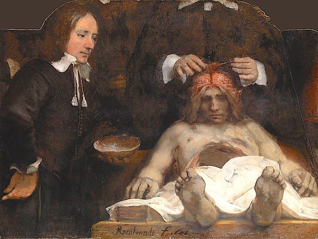
Twenty four years later, succeeding Tulp as praelector, Johannes Deijman (1620–1666) was the subject of Rembrandt’s second anatomy lesson: Anatomical Lesson of Dr. Deijman (1656) (Fig 2) displayed at the Hermitage, Amsterdam. Deijmann was born in Amsterdam in 1620. He studied medicine at Leyden and graduated MD in 1642 in Angers. He became Praelector in Amsterdam and died there in 1666.
It is a brain dissection ideally suited to Rembrandt’s method of darkening almost all of a scene while highlighting the key focal point. In this case your eyes are drawn to the victim’s skull as he lies motionless. His assistant Gijsbert Calkonen stands to the left, holding the section of skull that has been removed.
The original painting was reconstructed after Rembrandt’s later sketch for the design of its frame. This showed portraits of eight spectators, Dr. Deijman, his assistant, and the cadaveric “Black Jack” Joris Fonteijn, who was hanged for his “habitual criminality.” In contrast to the Tulp painting, the dissection of the abdomen has been completed, the thorax is unopened, and Deijman has removed the skull and lifted the falx and dura to display the cortical gyri. The painting closely represents the brain at dissection. It resembles Plate 67:2 of Vesalius’s De Fabrica (1543). By demonstrating the falx (sickle or scythe), which was the tool used by “Death” to cut down human life, the viewer is reminded of mortality, Memento Mori.
Artistically, Rembrandt’s Dr. Deijman is sometimes thought to be an even greater masterpiece than his Tulp. The foreshortened body is particularly dramatic, leading the viewer’s eye from the feet, across the open belly to the brain—reminiscent of the foreshortening in Mantegna’s Lamentation of Christ, which Rembrandt knew. The eyes stare directly at the viewer. The tone and contrast of the toes, lips, and skin and the rigor mortis are perhaps more vivid than in earlier paintings of Anatomy Lessons. When Sir Joshua Reynolds, the great English painter and critic saw the full original in 1781 he commented:
There is something sublime in the character of the head which reminds me of Michelangelo; the whole is finely painted, the colouring much like Titian.
Frederik Ruysch (1638–1731)
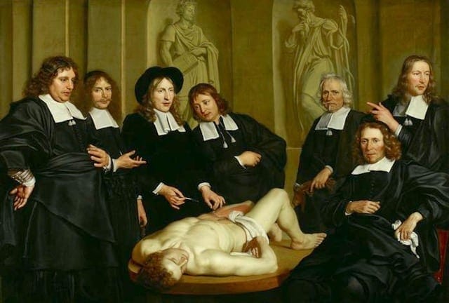
Though not painted by Rembrandt, the Dutch physician, anatomist, and botanist Frederik Ruysch was the subject of two important paintings in this series of “Dutch anatomy lessons.” Ruysch had succeeded Johannes Deijman as praelector anatomiae of the Amsterdam surgery guild in 1667. He retained this office until he died in 1731. The anatomy lesson of Professor Frederik Ruijsch (Fig 3) was painted by Adriaen Backer in 1670; it is housed in the Amsterdam Historical Museum. The corpse appears unopened; there are six spectators and Ruysch, who holds no visible dissecting instruments. The second painting by Johan van Neck in 1683 shows Ruysch dissecting the belly of a baby with five onlookers and (for reasons not immediately obvious) a youth with a baby’s skeleton.
Ruysch was a fine progressive anatomist. In his Opera Omnia he described a case of spina bifida with hydrocephalus, and he named ten cranial nerves compared to Vesalius’s seven. He developed techniques of injection of blood vessels with liquid wax before the dissection; this showed the “the bark-like substance of the brain consists merely of joined blood vessels,” thereby disproving Malpighi’s belief that it consisted of glands. Ruysch’s dissections of infants were carefully prepared to evoke the beauty of “innocents” dead before their time.
His legacy was a huge collection of anatomical preparations, bought by Tsar Peter the Great and displayed at the Kunstcamera Museum in St. Petersburg. In 1715 Ruysch was elected Fellow of the Royal Society and continued to teach anatomy until the age of eighty-five.
Comment
The anatomy lesson portraits were part of the Dutch Golden Age of art. The artists of this period included Rembrandt, the Delft master Johannes Vermeer, the landscape painter Jacob van Ruisdael, and Frans Hals. Rembrandt’s anatomy paintings demonstrate selected anatomical structures in a dissected body. But other Amsterdam anatomy lessons were more a record of a historical event for the guild and its proponents than a demonstration of detailed anatomical dissection. The regions dissected in different paintings are varied. Rembrandt’s mastery of the interplay of light and shade with strong contrasts (chiaroscuro) produced scenes of tension and drama. His unmatched artistic portraits showed how important were the leading praelectors of the guild.
As a sequel to this tradition, in 1925 The anatomy of Professor Louis Bolk was commissioned by Amsterdam anatomist Louis Bolk, to be painted by Martin Monnickendam. Bolk is surrounded by former pupils gathered around a cadaver of an orangutan, placed on a console—a portrayal of ambiguous meaning.
These great Dutch artists established a two-way link relating art and anatomy, which was displayed subsequently by other anatomists and pathologists including Charles Bell, who were to illustrate their own work and studies. Though perhaps essentially a display of the grandeur of this annual event, they also disclosed a more subtle, abstract interplay of art with life, death, and the dissemination of knowledge. Their portrayals of criminals’ corpses may overlay Shakespeare’s sentiment of “an insubstantial pageant faded, a rack left behind.”
JMS PEARCE, MD, FRCP, is a retired Consultant Neurologist and author.