
JMS Pearce
Hull, England
Letters, however small and dim, are comparatively large and distinct when seen through a glass globe filled with water.1
Seneca (c. 4 BC – AD 65)
The Dutch spectacle maker Zacharias Janssen (1585–c. 1632) and his father Hans are thought to have made one of the earliest (c. 1600) compound microscopes, which had both a convex and a concave lens mounted in a tube. It provided magnification probably twenty to thirty times normal size. Higher quality, hand-ground lenses could magnify up to 200 times. In the 1660s, Antonie van Leeuwenhoek (1632–1723) from Delft made simple one-lens microscopes using small, high-quality lenses that he ground himself with remarkable skill, patience, and compelled by a curiosity to see into a new microcosmic world.
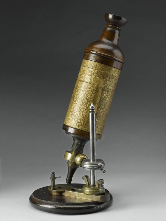
Leeuwenhoek observed animal and plant tissues, protozoa, human sperm and red blood cells, minerals, fossils, and many other “animalcules.”2 He presented his findings to the Royal Society in London, whose curator Robert Hooke was also making new observations with one of the earliest microscopes (made by Christopher Cock) (Fig 1). There can be no discovery more important than the cell, the unit of life, fundamental to all biology and medicine.3 Hooke was the first person to observe the cells of Quercus suber, the cork tree (which contained no nuclei),4 and he described them in a brilliant monograph, Micrographia, in 1665.
[T]hese pores, or cells…were indeed the first microscopical pores I ever saw, and perhaps, that were ever seen, for I had not met with any Writer or Person, that had made any mention of them before this… These pores, or cells [in cork] were not very deep, but consisted of a great many little Boxes, separated out of one continued long Pore.5
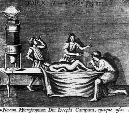
In the seventeenth century, the Jesuit Egyptologist Athanasius Kircher used first a simple, then a compound microscope, and saw “little worms” in the blood of patients suffering epidemic plague. Marcello Malpighi (1628–94) described microscopically the renal glomerulus (Malpighian corpuscle). The earliest picture, illustrating the use of the microscope in medicine, is from an engraving in Acta Eruditorum (1686), p. 372, titled “Novum Microscopium Dn Josephi Campani, ejusque usus,” reproduced by Singer.1 It shows a figure examining a leg ulcer with a microscope by the light of a candle; an enlargement of the microscope is shown on the left (Fig 2). Descartes too described his microscope, a modified telescope, in 1637.
Essays on the microscope (1787) was an early two volume text by George Adams. However, in the early 1800s, the preeminent histologist Xavier Bichat (1771–1802) found the early microscopes unsatisfactory because image blurring (spherical aberration) and color separation (chromatic aberration) hindered details of the microscopic images. Thus, many biologists refused to use a microscope.
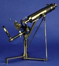
To correct these optical faults, in 1830 Joseph Jackson Lister with the instrument maker William Tulley made one of the first achromatic microscopes. Lister made these improvements in his spare time while fully engaged in his wine business.
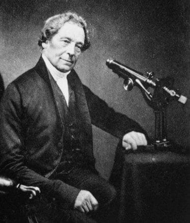
Joseph Jackson Lister FRS (1786–1869)
Joseph Jackson Lister’s (Fig 3) claim to fame lies in his often-forgotten improvement of the early microscope. The deserved reputation of his son, also Joseph, one of the greatest pioneers of surgery, has overshadowed his father’s works. Lister senior was born in Lothbury, London, son of Mary and John Lister, both members of the Society of Friends. At the age of fourteen Lister left boarding school in Dorset to work in the family wine business, living above the premises.
Joseph was an adroit amateur microscopist who, frustrated by the poor resolution of existing instruments, in his spare time designed and made achromatic lenses. He combined crown and flint glasses of different dispersion, an achromatic doublet, which minimized chromatic aberration by bringing red and blue light to the same focus, enabling greatly improved optical resolution. The use of microscopes then rapidly increased. Mirrors were added to enhance the light source and improve the image.
Lister had perfected the aplanatic-achromatic objective lens which James Smith incorporated into his “large best microscope.”6 He also worked with Andrew Ross assisting him in the production of achromatic objective lenses. His paper with Thomas Hodgkin (who described lymphoma) published in 1827 first described the morphology and accurate measurement of the red blood cells. His illustrious son Joseph wrote a fine account of this work.7 Lister published his optical work in a paper submitted to the Royal Society in 1830.8 Two years later he was elected Fellow of the Royal Society in recognition of his work on optics.
The surgeon and naturalist John Quekett FRS was a skilled microscopist who dedicated his Practical Treatise on the Use of the Microscope (1848) to Lister.9 Alexander Conrady (1866–1944), an optical designer and academician, posthumously transcribed Lister’s paper On the limit to defining-power, in vision with the unassisted eye, the telescope, and the microscope.10 It explained mathematically the optical resolution of the microscope’s objective lens.
On a visit to Ackworth School in West Yorkshire, he met Isabella Harris, a teacher, whom he married in 1818. They bought the grand Upton House, West Ham, where he lived for the rest of his life. Photographs show JJ Lister as a man of strikingly handsome appearance. He was much affected by the early deaths of his sons John and William Henry; and his wife Isabella died in 1864. His last years were lonely, lightened only by letters from Joseph in Edinburgh. He died in October 1869 at Upton House and was buried with Isabella in the Friends’ Burial Ground, Stoke Newington, Middlesex.
Owing to the scientific importance of the microscope in the 1830s, cells and cell theory became the focus of medical and biological research.11 Between 1838 and 1839, two German scientists, Mathias Schleiden (1804–81) and Theodor Schwann (1810–82), proposed cell theory (Theorie der Zellen): that cells are the fundamental structural and functional units of plant and animal life.12 Schwann’s original ideas were taken up by Rudolf Virchow (1821–1902), the most influential pathologist of the 1800s.
Much later came the refinements of the Scanning Electron Microscope (1947) which uses a focused beam of electrons to scan the surface of a sample; confocal microscopes using point illumination of fluorescent images; and the Transmission Electron Microscope that uses a beam of accelerated electrons rather than light to magnify times 10,000 to 10,000,000.
References
- Singer C. Notes on the Early History of Microscopy. Proceedings of the Royal Society of Medicine 1914;247-79.
- Dunea G. Van Leeuwenhoek’s discovery of “animalcules.” Hektoen International Fall 2018.
- Gest H. Homage to Robert Hooke (1635–1703): New Insights from the Recently Discovered Hooke Folio. Perspectives in Biology and Medicine 2009;52(3):392-9.
- Pearce JMS. Robert Hooke and Micrographia. Hektoen International Spring 2023.
- Hooke R. Micrographia. Published by the Royal Society, 1665. British Library. https://bl.uk/collection-items/micrographia-by-robert-hooke-1665.
- Sobel B. de Groot J. “Large Best” Microscope by James Smith. Microscope-Antiques.com. https://microscope-antiques.com/smith22.html.
- Lister J. Lister, Joseph Jackson. Dictionary of National Biography vol. 33, 1885-1900.
- Lister JJ. On the Improvement of Achromatic Compound Microscopes. Philosophical Transactions of the Royal Society 1830;120:187-200.
- Allen E, Turk JL. Microscopes in the Hunterian Museum. Annals of the Royal College of Surgeons of England 1982;64:414-18.
- Lister JJ. On the Limit to Defining-power, in Vision With the Unassisted Eye, the Telescope, and the Microscope. Journal of the Royal Microscopical Society 1913;33:34-5.
- Science Museum. The Microscope. August 19, 2019. https://www.sciencemuseum.org.uk/objects-and-stories/medicine/microscope.
- Pearce JMS. The Beginnings of Cell Theory: Schleiden, Schwann and Virchow. Hektoen International Summer 2022.
JMS PEARCE is a retired neurologist and author with a particular interest in the history of medicine and science.
Highlighted in Frontispiece Volume 15, Issue 4 – Fall 2023