JMS Pearce
Hull, England
 |
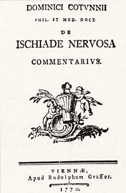
| Fig 1. Dominici Cotugno’s De Ischiade Nervosa, 1764. 1770. |
Access to cerebrospinal fluid (CSF) in life as an aid to diagnosis proved impossible until lumbar puncture. Galen of Pergamon (AD 130–200) failed to recognize CSF; he described a vaporous, not aqueous, humor that he called περιττώματα (residues) in the cerebral ventricles. Cerebrospinal fluid in the ventricles was probably first shown by the somewhat histrionic Venetian physician and anatomist Nicolò Massa in 1536: “semp has cavitates inveni plenas, aut semiplenas dictae aquose substantiae.” [I have said, always these cavities are full, or half full of the so-called watery substance.]1 The anatomist Vesalius in De humani corporis fabrica (1543) also described a watery fluid surrounding the brain and present in the ventricles.
CSF in the spinal canal was not shown until 1692 when Valsalva cut a dog’s spinal cord and observed a clear, watery liquid resembling synovial fluid. CSF was observed by Emanuel Swedenborg between 1741 and 1744 in a manuscript not published until 1887, but mentioned by Albrecht von Haller in his textbook of 1762. In hydrocephalic patients, von Haller also described vapors, not fluid, in the ventricles, but watery tumors [swellings] in the lower part of the spinal marrow.
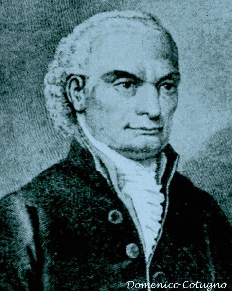
In 1764, Domenico Cotugno (Dominic Cotunnii, 1736–1822) of Ruvo di Puglia, gave the definitive description of cerebrospinal fluid (liquor Cotunnii) enveloped by membranes of the cord2,3:
all that space which is between the Vagina of the Dura Mater, and the spinal marrow, is always found to be filled…with water, like that which the Pericardium contains or such as fills the hollows of the ventricles of the brain, the labyrinth of the ear or other cavitiesi of the body, which are impervious to the air.4,5
To prove the free circulation between the cranial and spinal dura, Cotugno stood cadavers on their feet and decapitated them to observe the flow of CSF.
We now know that the epithelial cells of the choroid plexuses secrete cerebrospinal fluid by a mechanism that involves transport of sodium, chloride, and bicarbonate ions, which creates an osmotic gradient driving the secretion and absorption of CSF betwen the blood and the ventricles. But Cotugno’s work achieved little recognition until republished by Magendie in 1827. Amongst several other original discoveries he demonstrated the aural labyrinths filled with fluid; and simultaneously with Scarpa he discovered the nasopalatine nerve. He gave a classic account of sciatica, which he attributed to a distention of the “vaginae” of the ischiadic [sciatic] nerves. In this text he gave his account of cerebrospinal fluid. He also was the first to describe intestinal ulcers in typhoid fever. He also observed that urine precipitated on boiling in acute nephritis.
Diagnostic lumbar puncture
 |
|
Fig 2. Domenico Cotugno. Via Wikimedia. Public domain. |
Aspiration of the CSF was first used as a therapeutic procedure in meningitis before the advent of antibiotics.6 A different use by the New York physician Leonard Corning in 1885 was for spinal anesthesia. Examination of the CSF by lumbar puncture was soon employed to establish the diagnosis of many diseases of the brain, spinal cord, and nerve roots. By 1906 Peter Marshall was able to write: “Lumbar puncture is now a well-established aid to the diagnosis of certain diseases, and deserves to rank with paracentesis of other body cavities in the diagnostic armature of the physician. It has not so far, however, acquired the position among the rank and file of practitioners which its importance merits.”7
Though Quincke’s name is rightly attached to the procedure of lumbar puncture, Walter Essex Wynter (1860–1945) in 1889, the same year, devised a comparable if cruder technique for therapeutic aspiration of CSF8 performed through a small bore rubber (Southey’s) tube.
While a registrar at the Middlesex Hospital, Wynter reported in The Lancet of 1891 aspiration of CSF in four children with meningitis.9 In one it was the sequel to an ear infection, in the other three it was tuberculous. Wynter made a small incision at L2, cut down to the dura, then inserted a Southey’s tube with a rubber drainage to withdraw the infected fluid and reduce the pressure. The procedure afforded but short-lived relief and all four patients died. (Southey’s tubes were still in occasional use in 1960 to relieve gross dropsy in the legs, which were left dependent overnight to drain liters of edema fluid into a large bucket.)
Wynter was educated at Epsom College, Surrey, and the Middlesex Hospital. His father Andrew Wynter, a general practitioner, edited the British Medical Journal (1855–61). Walter Wynter was appointed a physician to the Middlesex Hospital in 1901.
Heinrich Irenaeus Quincke (1842–1922)
 |
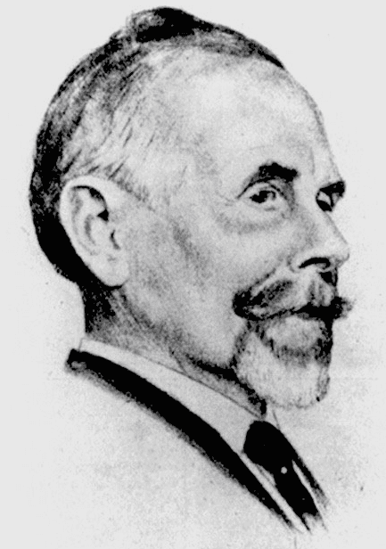
| Fig 3. Heinrich Irenaeus Quincke. From Minagar A, Lowis GW.13 |
Diagnostic puncture through a needle was first performed in the same year by Heinrich Irenaeus Quincke at Kiel.10 Much influenced by his teacher Friederich Frerichs in Berlin, in 1872 Quincke demonstrated the flow of CSF in the subarachnoid space in dogs and rabbits by injecting red sulphide of mercury into the CSF.
Twenty years later, whilst pondering the cause of headaches associated with hydrocephalus, he reasoned that the removal of some CSF might reduce its production or increase its absorption. One month before Wynter’s Lancet paper (which he acknowledged), Quincke reported his first lumbar puncture in Ueber hydrocephalus to the Tenth Congress of Internal Medicine at Wiesbaden.11
Case 1 was a boy of 12 years who died despite six punctures of the ventricles through a trephine, performed in 1888. Case 2 was a boy aged one year nine months, comatose with suspected tuberculous meningitis. He performed three lumbar punctures at three-day intervals in December 1890:
I punctured the subarachnoid space in the lumbar area, passing a very fine cannula 2cm. deep between the third and fourth lumbar spinal arches and drop by drop I drained a few cubic centimetres of watery fluid… one could see clearly increases with expiration and decrease with inspiration.
The child recovered and the nature of the meningtis or meningism remains unknown. Case 3 was a man aged twenty-five with chronic hydrocephalus, who suffered from severe headaches. These were relieved by lumbar puncture, but again the aetiology was not established.8
His paper12 in September 1891 described lumbar puncture in five children and five adults. He had measured pressure at the start and end of the procedure, and noted the raised protein and low sugar content in the CSF in purulent meningitis. He demonstrated tubercle bacilli (first shown by Koch in 1882) in the CSF in tuberculous meningitis.
Heinrich Irenaeus Quincke13 was born in Frankfurt-an-der-Oder, the son of a Berlin physician and a Huguenot mother. He read medicine at Berlin, Wurzburg and Heidelberg, graduating MD in 1863. He was later appointed professor of medicine in Berne and then in Kiel.
He established Quincke’s triad: jaundice, melena, and upper abdominal pain, which suggests bleeding from the biliary tree (hemobilia), a rare but often iatrogenic syndrome. He advocated surgery for lung abscess and observed the angioedema of anaphylaxis, referred to as Quincke’s edema. He observed the pulsation of nailbed capillaries in aortic regurgitation, Quincke’s sign.
In 1909, Quincke was nominated for the Nobel Prize in Physiology or Medicine for his 1891 work on lumbar puncture. It was however rejected because eighteen years had elapsed between the date of the discovery and the nomination. Sic transit gloria mundi.
Queckenstedt’s test
An important test related to lumbar puncture was devised in 1916 by Hans Heinrich Georg Queckenstedt of Leipzig to detect spinal canal obstruction. He described:
“The narrowed [spinal] channel impedes movement of fluid with an increase in pressure above the compression site…. The increment in pressure above the obstruction can be demonstrated by compression of the neck…, which produces an increase in venous blood in the cranial cavity, with concomitant reduction in space for the cerebrospinal fluid…. The increased fluid pressure immediately transmitted throughout the system normally can be demonstrated with …a manometer attached to a lumbar puncture needle. In lesions of the cord the manometric change is greatly retarded.”14
More recently, spinal imaging has made obsolete this previously invaluable test.
References
- Massa N. Liber Introductorius Anatomiae, 1st ed. Venetio: Francesco Bindoni et Masseo Pasini, 1536. cited by Herbowski L. “The Maze of the Cerebrospinal Fluid Discovery.” Anatomy Research International vol. 2013, Article ID 596027, 8 pages, 2013.
- Cotunnii D. De Ischiade nervosa commentarius. Neapoli: Simoniana 1764, and in English, London: J Wilkie 1775.
- Pearce JMS. “Cotugno and cerebrospinal fluid.” J Neurol Neurosurg Psychiatry. 2004;75(9):1299.
- Viets HR. “Domenico Cotugno: his Description of the Cerebrospinal Fluid, with a translation of part of his De Ischiade Nervosa Commentarius (1764) and a bibliography of his important works.” Bulletin of the Institute of the History of Medicine 3, no. 9 (1935): 701–38.
- Levinson A. “Domenico Cotugno.” Ann Med Hist. 1936;8(1):1-9.
- Frederiks JA, Koehler PJ. “The first lumbar puncture.” J Hist Neurosci. 1997;6(2):147-53.
- Marshall P. “A Clinical Investigation of Lumbar Puncture.” Edinb Med J. 1909;3(3):231-255.
- Pearce JMS. “Walter Essex Wynter, Quincke, and lumbar puncture.” J Neurol Neurosurg Psychiatry. 1994; 57(2):179.
- Wynter WE. “Four cases of tubercular meningitis in which paracentesis was performed for the relief of fluid pressure.” Lancet 1891;1:981-2.
- Stewart P. “The Clinical Significance of the Cerebro-Spinal Fluid.” Edinb Med J. 1906;19(5):429-443.
- Quincke HI. “Ueber hydrocephalus.” Verhandlung des Congress Innere Medizin (X), 1891;321-39.
- Quincke HI. “Die lumbalpunction des Hydrocephalus.” Berl Klin Wochenschr 1891; 28:929-33.
- Minagar A, Lowis GW. “Dr Heinrich Irenaeus Quincke (1842–1922): Clinical Neurologist of Kiel.” Journal of Medical Biography 2001;9(1):12-15.
- Queckenstedt HHG. “Zur Diagnose der Rückenmarkskompression.” Deutsche Zeitschrift für Nervenheilkunde, 1916, 55: 325-333. Cited in Talbott JH. A Biographical History of Medicine: excerpts and essays on the men and their work. New York: Grunne and Stratton 1970; p. 983.
JMS PEARCE is a retired neurologist and author with a particular interest in the history of medicine and science.

Leave a Reply