
JMS Pearce
Hull, England, United Kingdom

The eminent physician Johannes Jakob Wepfer (1620-1695) was born in Schaffhausen, Switzerland, on the right bank of the Rhine. Little is written of his early years but the child Wepfer may have gazed and wondered about Schaffhausen’s countryside, its many oriel windows, and the rounded Munot fortress designed by Albrecht Dürer. His father Georg was a merchant and local Councilor, whose family from the Canton Thurgau contained doctors, burgomasters, guildmasters, and judges.
After studying in Strasburg, he graduated in Basle under Caspar Bauhin and Felix Plater, and in Padua with Thomas Bartholin. For his MD Wepfer published De palpitatione cordis (Basel, 1647), which stoutly supported Harvey’s then controversial ideas on the circulation of blood. After studies abroad, he returned to his native town where he built a successful practice and was appointed physician to the Palatinate Elector Karl in 1685.1 In 1648, he became municipal physician of Schaffhausen. His practice spread through Switzerland and Germany. He became physician of the cloister at Rheinau and consultant to several German princes.
His name was hallowed as “The Hippocrates of Helvetia.”2 A man of temperate habits, he was an industrious scholar, devoted to reading and the scriptures.
Apoplexy
His mind was active, ambitious, and adventurous. Keen on experimental physiology and anatomy, he obtained permission of the government to start routine autopsies. From these he showed that apoplexy was caused by cerebral hemorrhage.3 In Historiae apoplecticorum, published in 1658 is a detailed description of four such cases, the first studied in 1655.4
In one case: “. . . cut the dura mater, much blood discharged from the space between it and the pia-mater [subarachnoid space] . . .” in another case: bleeding with ruptured anterior cerebral artery branch; in one more case, with serous liquid accumulation.5
A later edition contained the history of the cerebral hemorrhage and autopsy of Marcello Malpighi, who was the Pope’s physician and author of De viscerum structura exercitatio anatomica, London 1669:
“On July 25th 1694 at which Time he was seized in the 66th year of his Age, about 1 a clock in the Afternoon, with an Apoplexy . . . attended with a Palsie of the whole right Side, and a distortion of the mouth and Right Eye.”
A second episode on November 29th proved fatal. Wepfer reported:
“[within] the Cavity of the right Ventricle of the Brain an Extravasation of about 2 pints of black clotted Blood, which was the cause of his Apoplexy and Death . . .”
Six years before Thomas Willis’s description of his arterial circle, Wepfer in 1658 had described the vessels of the dura mater, the vertebral and internal carotid arteries with their branches and their terminal ramifications.5 Wepfer’s description (Fig 2) clearly shows that he had fully identified the vessels that make up the circle, though he gave no illustration. His account was more accurate and detailed than the earlier ones of Fallopius in his Anatomical Observations (1561), Giulio Casserio’s Tabulae Anatomicae (1632), and Johann Veslingus’s Syntagma Anatomicum (1641) (Fig 3).6
He also described a stroke caused by carotid occlusion. His book Observationes Medico-practicae de Affectibus capitis… scaphasii included 222 neurological patients. Isler reported its descriptions of basilar artery migraine, a migrainous stroke in a child, and the visual aura of migraine. 7
Wepfer recognized the association and mechanism of polyuria with “extremely small and slender kidneys” 150 years before Richard Bright’s 1827 account of dropsy and albuminuria in what was subsequently shown to be mesangiocapillary (membranoproliferative) glomerulonephritis. Wepfer investigated the toxicology of water hemlock, prompted by a local village poisoning. He studied the toxicology of mercury, arsenic, antimony, nux vomica, and ergot. Two years before Malpighi’s De hepate 1666, he described the lobular structure of the pig’s liver, distinguishing its venous and biliary systems.

Aged seventy-five, he died in 1695 of severe aortic sclerosis, which he diagnosed himself. The post-mortem findings were published in his biography Memoria Wepferiana in 1727.
from the aortic valve . . . all the way to the very iliac ramifications themselves, were deposits of semi-circular shape whose consistency varied from gristle to frank bone.
Both his son Johann Conrad and his grandson Georg Michael were physicians. His youngest daughter married his friend Johann Conrad Brunner (1653-1727), the much celebrated Swiss physiologist who experimented on pancreatic functiona and showed the secretory (Brunner’s) glands in the duodenum.
The Medical Faculty of Leyden bought the medico-literary memoirs of the Wepfer family in 1774 for one hundred and twenty gold ducats.
End notes
- Brunner reported thirst, polyuria, and increased appetite in panceatectomized dogs, but failed to connect this to human diabetes.
References
- Fischer H. Johann Jakob Wepfer, 1620-1695: ein Beitrag zur Medizingeschichte des 17. Jahrhunderts, Zurich, 1931.
- Eichenberger P. Johann Jakob Wepfer (1620-1695) als klinischer Praktiker, by (Basler Veroffentlichungen zur Geschichte der Medizin und der Biologie, Fasc. XXVI), Basleand Stutgart, Schwabe, 1969.
- Pearce JMS (1997). Johann Jakob Wepfer (1620-1695) and Cerebral Haemorrhage J. Neurol Neurosurg Psychiatry. 62,387.
- Wepfer JJ Historiae apoplecticorum. (1658). English translation from Bagvili’s Practice of Physik, London 1704: cited in Major RH. Classic descriptions of disease, 3rd ed, Illinois: Charles C Thomas. pp. 474-7.
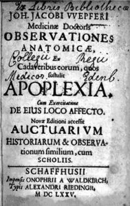
- Wepfer J J. Observationes anatomicae ex cadaveribus eorum quos sustulit apoplexia… Schaffhausen: O a Waldkirch; 1658. [From: http://books.google.com].
- Meyer A, Hierons R. (1962) Observations on the history of the ‘Circle Of Willis’. Med Hist ;6:119-30.
- Wepfer J. Observationes Medico-practicae de Affectibus capitis… scaphasii. JA Ziegler Schaffenhausen. Obs. CVIII, 480. cited by Isler H. In: Blau JN (Ed.) Migraine. London: Chapman Hall, 1987:665.
- Karenberg A. Historic review: select chapters of a history of stroke. Neurol Res Pract. 2020;2:34.
- Dumitrescu AM, Costea CF, Cucu AI, et al. The discovery of the circle of Willis as a result of using the scientific method in anatomical dissection. Rom J Morphol Embryol. 2020;61(3):959-965.
JMS PEARCE, MD, FRCP, is emeritus consultant neurologist in the Department of Neurology at the Hull Royal Infirmary, England.