Arpan K. Banerjee
Solihull, United Kingdom
Francis Henry Williams was born in Massachusetts on July 15, 1852. His father was a professor of ophthalmology at Harvard Medical School. Williams graduated in chemistry in 1873 from the Massachusetts Institute of Technology and in medicine from Harvard in 1877.1 He followed this with two years of training in Vienna and Paris. His early research was in infections, in particular the use of diphtheria antitoxin.
Although Williams started off as a physician, the discovery of X-rays by Rontgen fired his interest in its potential applications. In 1896 he reproduced Rontgen’s experiments in the laboratory of physics at the Massachusetts Institute of Technology, which was headed by Charles Cross.2 Before Boston City Hospital had its own department of radiology, patients were transferred here to have X-rays taken in the evenings. In 1896 Williams opened a radiology unit in the basement of Boston City Hospital and X-rayed patients from Harvard, Tufts, and Boston Universities.
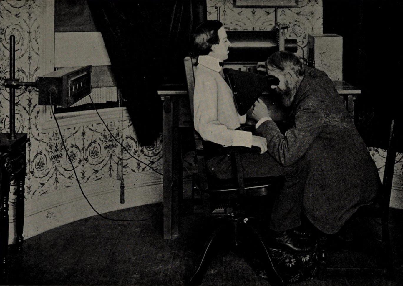
In the early days, chest X-rays required a long exposure time. Because of this, Williams preferred to do fluoroscopic real-time examinations on his patients, which showed the chest in more detail and more clearly than on plain film studies. He gained prodigious experience in the examination of the chest in a relatively short time, and by 1897 had produced over 409 volumes (each of around 250 pages) of drawings of patients with chest diseases. He was one of the earliest radiologists to describe the apical changes on chest X-ray in patients with tuberculosis. He also described other chest X-ray findings such as emphysema and pleural effusions.


In 1899 he performed early fluoroscopic studies on the gastrointestinal tract and stomach with his fellow physiologist Walter Cannon (1871-1945), who coined the term “fight or flight response” and later became a famous professor of physiology at Harvard (1906-1942).
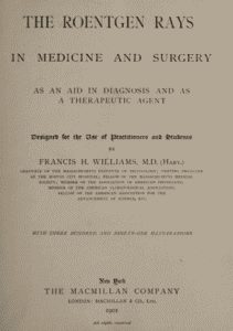
In 1901 Williams wrote The Roentgen rays in medicine and surgery, which boasted 391 illustrations.3 He described changes on chest radiographs in a variety of medical conditions such as pneumothorax, pleural effusion, heart failure, and emphysema. He also described the appearance of a thoracic aortic aneurysm in 1898 and even described calcification within the heart.
Williams remained an astute clinician as well as being a brilliant scientist and investigator. He became interested in the therapeutic properties of X-rays, as was common with the early pioneers, and published Radium treatment of skin diseases, new growths, diseases of the eyes and tonsils.4 He was also aware of the harmful effects of X-rays and was an early advocate of radiation protection.5
He became President of the Association of American Physicians from 1917-18 and an honorary member of the American Rontgen Ray Society and the Radiology Society of North America. He died on June 22, 1936 at the age of eighty-three. An eponymous lecture is still delivered annually at Boston University.
References
- Greene R. “Francis H Williams.” Radiographics 1991, 11, 325-332.
- Williams FH. “Notes on X-rays.” Boston Med Surgical Journal 1896 April 30.
- Williams FH. The Roentgen Rays in Medicine and Surgery. 1901: MacMillan.
- Pendergrass EP. “Editorial: FH Williams.” Radiology 1953, (60) 5 May1.
- Nath H. “FH Williams: The Father of Cardiac Fluoroscopy.” American J Radiology 1993, (160) 260.
ARPAN K. BANERJEE, MBBS (LOND), FRCP, FRCR, FBIR, qualified in medicine at St. Thomas’s Hospital Medical School, London. He was a consultant radiologist in Birmingham from 1995-2019. He served on the scientific committee of the Royal College of Radiologists 2012-2016. He was Chairman of the British Society for the History of Radiology from 2012-2017. He is Chairman of ISHRAD and adviser to radiopaedia. He is the author/co-author of numerous papers and articles on a variety of clinical medical, radiological, and medical historical topics and seven books including Classic Papers in Modern Diagnostic Radiology 2005 and “The History of Radiology” OUP 2013.

Leave a Reply