

Paris during the greater part of the nineteenth century was the mecca of medicine, home of great surgeons and great physicians. Doctors from all over the world flocked to its hospitals to learn from its famous professors and study pathology in their amply supplied dissecting rooms.
Among these students was a Scottish physician named Robert Carswell. He was a talented young man who could draw sketches and paint in watercolor. Born in 1793, he attended the faculty of medicine in Glasgow, and after being recognized as a skillful illustrator was hired by a practicing Edinburgh physician to make a collection of anatomical drawings for his lectures on the practice of physic. After graduation he went to Paris, and also to Lyon, spending two years painting and drawing in hospitals and mortuaries. In 1826 he obtained an MD degree from Aberdeen, then returned to Paris to work with the celebrated physician Pierre Louis.1 By the time he left Paris he had completed over 1,000 pictures.
At age thirty-five Carswell became the first professor of pathological anatomy at University College London (1828). He also received a hospital appointment, but his lack of success in private practice and his poor health led him to resign these appointments in 1840. Being fluent in French, he was appointed physician to Leopold I, uncle of Queen Victoria and King of the newly created kingdom of Belgium. Later, he treated the former French king Louis Philippe, who lived in exile in London, and for this was knighted by Queen Victoria. For the rest of his life he lived mostly in Laeken, near Brussels, and died of chronic lung disease in 1857.
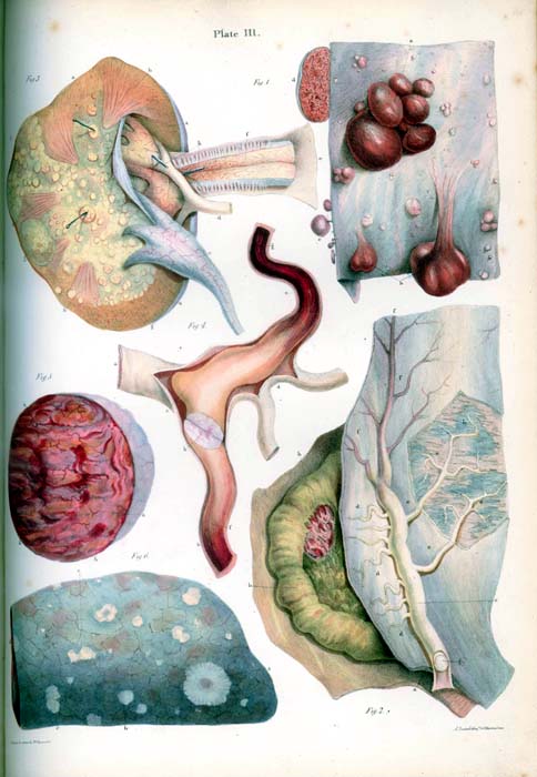
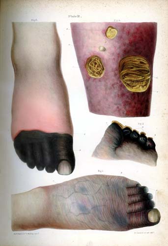
In 1838 Carswell published a beautifully illustrated atlas of drawings and watercolor paintings. The entire collection of his work comprises 1,034 representations of pathology specimens of every system of the body, and includes pictures of pericardial disease, syphilitic aortic aneurysms, aortic stenosis, tetralogy of Fallot, coronary arterial sclerosis, and Hodgkin’s disease, as well as ninety-nine images of the brain and spinal cord. Carswell was one of the first to illustrate the cerebral lesions of multiple sclerosis—in a patient he never saw but was told suffered from paralysis. He also described the “fatal black tumor” for which he coined the name melanoma. He was an important illustrator, living as he did before the introduction of microscopy and photography into medicine.1

Reference
Hollman A. The paintings of pathological anatomy by Sir Robert Carswell (1793-1857). Br. Heart J 1995;74:566.