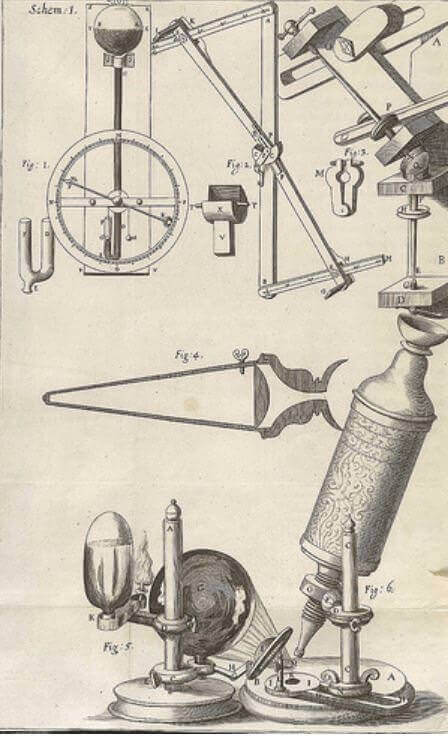
Marcello Malpighi was fortunate to live at a time when microscopes of sufficient power became available for scientific studies, culminating centuries of attempts to use the optic properties of glass to magnify the image of objects. Such efforts go back at least to the Romans, who for this purpose ground glass into the shape of lentils, hence the term lenses. It is believed that spectacles were invented sometime in the thirteenth century. Around 1590 the Dutch spectacle makers Janssen built a rudimentary microscope by inserting several lenses into one tube. Further progress ensued, and by the middle of the seventeenth-century microscopes of sufficient power enabled Robert Hooke, curator of experiments and fellow of the newly formed Royal Society, to describe the constituent building blocks of plants as “cells”—by analogy with beehives or the cells in which monks slept in monasteries. Also around 1660 in the Netherlands, Antonie von Leeuwenhoek built a microscope of sufficient magnification to see muscle fibers, bacteria, spermatozoa, and red cells in capillaries—and called the living organisms animalcules. It was also at this time that Marcello Malpighi began his studies.
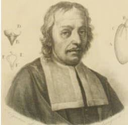
Born in 1628, Malpighi entered the University of Bologna at the age of seventeen, first studying philosophy and physics, but switching his interests to anatomy after being awarded a doctorate of medicine in 1653. In 1656 he was given a teaching appointment in Bologna, and except for temporary professorial appointments at Pisa and Messina he spent most of his life there. Using the newly developed tool of microscopy, he studied plants, legumes, and flowers, and also insects, frogs, higher animals, and humans.
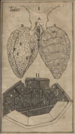
He looked at brains, liver, tongue, lung, bone, and skeletal muscle, describing their architecture and postulating what their function might be. In the mesentery of frogs he saw capillaries and also saw them in the lungs, where he also noted that the structure of the lungs consisted of alveoli. He was one of the first to describe red blood cells (as very small red particles), also the components of a blood clot, taste buds, and the ridges in the skin of fingers later used in fingerprinting for identification.
Using a microscope with x30 magnification and sometimes with prior dye injection, he described the renal glomeruli (now also named the Malpighian corpuscle), which when injected “turned black . . . hanging like apples from the blood vessels which, swollen with the black fluid, look like a beautiful tree.” He described the (Malpighian) pyramids in the renal medulla and the collecting ducts, and noted the opening of these ducts at the papilla. He also is credited with describing the (Malpighian) corpuscles in the spleen, and the (Malpighian) layer of mucus beneath the skin with its pigmented cells accounting for different colors of the skin itself.
He also built a reputation in botany and zoology. He studied silkworms and noted the (Malpighian) excretory tubule of insects, and even has genus of plants, the Malpighiaceae, named after him. At the peak of his reputation in 1691 he went to Rome at the invitation of Pope Innocent XII, becoming his personal physician, and teaching at the university. It was in Rome that he died at age sixty-sixty of a stroke. The medical community remembers him by the many anatomical structures named after him, and Bologna has raised a monument in honor of its illustrious adopted son.