
JMS Pearce
East Yorks, England
Home to the oldest western university,1 the University of Bologna was founded in 1088 and was a center of intellectual life during the Middle Ages, attracting scholars from throughout Europe. The University began as a law school. Medical teaching started circa 1156 and was taught in Latin translations, principally based on Arabic texts.
The Renaissance of anatomy did not occur overnight. Erasistratus (c. 325–c. 250 BC) and Herophilus (c. 330–c. 260 B.C.) from the Alexandrian school, and Rufus of Ephesus (c. 80–AD 150) were amongst the first scholars of anatomy before Galen (129–c. AD 210).i In the 13th century, medical teaching was theoretical with little clinical demonstration. Surgery had a lower status than medicine and its herbal remedies. For centuries there prevailed opposition by religious authorities to the growth of knowledge, whose possessors became “victims of calumny and persecution.”ii Gradually, advances were made at celebrated schools such as Bologna, Padua, and Salerno,iii well described centuries later by Hamilton.2 The crafts of surgery were improved by Roger of Salerno (1140–1195), also known as Roger of Palermo, whose 1170 Practica Chirurgiae was a classic for over 200 years.
At Bologna dissections were re-established by Mondino de’Luzzi (1270–1326) in the 14th century after the hiatus that followed the Alexandrian anatomists.iv Surprisingly, there was little general interest in biological science, natural history, or experimentation. The reason for dissection was mainly to obtain legal evidence for the cause of death. The oldest anatomical theatre was in Padua, founded in 1594 by Girolamo Fabricius Acquapendente (1533–1619); and in 1596 the Theatrum Anatomicum at Leiden was built.
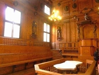
Anatomy theatres proliferated in universities across Europe. Amongst the oldest, Teatro Anatomico was in Bologna inside the Palazzo dell’Archiginnasio. It was built in 1637, carved in spruce wood, designed by Antonio Levanti, but not finished for many years. Although badly damaged during World War II it still resembles the ancient candlelit theatre. (Figs 1,2) This elliptical theatre has six tiers each with a clear view of the marble dissecting table for up to 300 spectators. The inscription, Hic locus est ubi mors gaudet succurrere vitae (This is a place where the dead are glad to succour the living) is still open to view. It has not been used since 1872 because the political disruptions of the Napoleonic era prevented public dissections and the study of anatomy.v However, the ancient theatre remained as a building with its original dissecting table and many surgical tools and specimens. From its inception it was repeatedly extended and modified.
Giovanna Ferrari describes this space:
The dissecting table was surrounded by a balustrade to protect it from the scholars who crowded round it. Three rows of benches and an aisle ran around all four walls. The anatomy professor’s cathedra was situated along one of the end walls, as were the prior’s chair and the seats for the counsellors. Against the opposite wall sat the authorities. Along the side walls, “on the right will sit the university, accompanied by its porters with the maces, and the notary; on the left, on the other hand, will sit the entire body of doctors, according to the seniority of their doctorates; and all around, the young scholars. Any seats left over were occupied by ordinary citizens. . . .vi

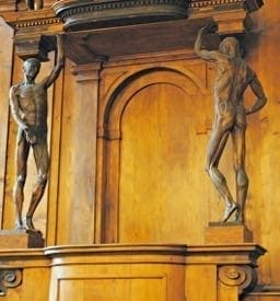
In 1723 the ruling authorities decided to redevelop the anatomy theatre because local benefactors offered to pay for six new statues. Domenico Gianotti, a celebrated wood-carver from Lucca, was engaged. He made life-sized figures of twelve ancient anatomists: Hippocrates, Galen, Mondino de’Luzzi, Varignana, Malpighi, Sbaraglia, Argelata, Aranzio, Varolio, Tagliacozzi, Bartoletti, and Fracassati. But Gianotti’s skills were criticized; he was more experienced with the draped figure than the nude, and skinning the nude was not for him to undertake.5 According to Professor Medici, “now there comes Ercole Lelli who of his own free will offered to carve in wood the two anatomical figures (Fig 3.) free of charge.”5
Ercole Lelli (1702–1766), at this time only 30 years old, had long been interested in both painting and studying the human figure, and had frequented dissecting rooms to familiarize himself with the bones and muscles. Lelli was a pupil of the painter Giovanni Pietro Zanotti. He began to make wax models of the viscera: “so lifelike nothing equal to them has yet been seen.” Starting in 1742 he began to prepare artistic anatomical displays at Bologna University. Lelli made eight display cabinets from contemporary wood and glass, restored after World War II; they contain his representations of the whole muscular apparatus. They are aligned along the right side of the corridor on the first floor of the Institute of Human Anatomy. The marble dissecting table used by the famous anatomist Luigi Galvani (1737–1798) is placed at one end of the corridor. From Lelli’s wax anatomical models the copies were carved in linden-wood so skillfully and beautifully that they were acclaimed masterpieces. In 1734 the theatre was completed.
The work greatly impressed the scholarly Cardinal of Bologna, Prospero Lambertini, who advised the Senate to create the Institute of Sciences’ Anatomical Museum to restore Bologna’s primacy in medical learning.vii Lambertini became Pope Benedict XIV and at his own expense in 1742 paid for the museum display for students of anatomy. Lelli was made custodian. In 1743 his talented assistant, the Bolognese sculptor Giovanni Manzolini assisted by a surgeon, Boari, came to excel his teacher as a modeller in wax. He too was excelled by his pupil and later wife, Anna Morandi Manzolini. Their effigies in wax are still preserved. Giovanni Manzolini resigned in late 1746; he believed that Lelli had traduced his greater knowledge of anatomy and sculpture.7
The surviving theatre is elaborately decorated. Carved statues of physicians such as Hippocrates and Galen stand in niches. Because in ancient times surgeons would consult the stars before operating, the ceiling is decorated with astrological symbols: protections by signs of the zodiac. It recalled the concept of man’s affinity with the cosmos. Lelli’s famous statues of the Spellati that are skinned anatomical models displaying the muscles beneath the skin (écorché) hold up the canopy above the teacher’s chair. (Fig 2.) In 1746 Lelli became a member both of the Bolognese art society, Accademia Clementina, and the Istituto delle Scienze. He also made several medals for the local mint. A few of his pictures and some engravings of Hagar and Ishmael still exist. In 1759 Lelli was appointed director of the Academy at Bologna, where he died. His book, Anatomia esterna del corpo umano, was published posthumously (1770) in Bologna. Between 1789 and 1815, the museum acquired 23 superb wax modelsviii made by Clemente Susini of Florence from 1782.ix
On January 29, 1944, during World War II, the theatre was destroyed. It was later skillfully rebuilt using original remnants recovered from the rubble.
Anatomists in Bologna
The University of Bologna began as a law school. Medical teaching was primarily theoretical with sparse clinical content. Yet it was here that dissections were established mainly to demonstrate the cause of death and perhaps to verify the Al-Qānūn fī al-ṭibb (The Canon of Medicine) based on Aristotelian philosophy and Arabic Medicine of the revered Avicenna (Ibn Sīnā) (980–1037), the Persian polymath.
The surgical school in Bologna slowly developed alongside the beginnings of anatomy. A noted exponent was William of Saliceto (?1215–?1280), the ablest Italian surgeon of the 13th century, who reintroduced the surgical knife to replace the Arabic method of burning with cautery. His treatise Chirurgia was original, expounding the principles of regional and surgical anatomy. He also devised techniques for nerve suture anastomosis. His contemporary at Bologna, Thaddeus Alderotti of Florence (c. 1223–1303) advanced anatomy by insisting on practical experience from dissection of the human body rather than studying books.2,8,9 Henri de Mondeville (c. 1270–1320) also studied in Bologna. In treating wounds he advocated cleanliness trying to prevent “laudable pus”; he aimed at healing by first intention, a new concept for this period. His skills spread to Europe when he moved to Montpelier.
At this time Mondino di Luzzi (c. 1275–1326), a pupil of Thaddeus, studied anatomy from his own public dissections. Creatively, he considered physiological mechanisms and their clinical application. His book Anathomia (1316, but not printed until 1478)x was the first illustrated manual of anatomy after Galen’s. It became a classical text with more than 30 editions.xi Di Luzzi improved dissecting techniques to show particular anatomical structures.xii Not until 1521 was further progress evident. In Bologna, Berengario da Carpi (?1466–?1530), the most important anatomist before Vesalius, produced even more detailed text and pictures guiding anatomy towards the science of observation.xiii Bold and original, he displayed a healthy skepticism of past dogma. He refuted Galenic ideas of a human rete mirabile, and Aristotle’s third ventricle of the heart. Many subsequent writers added fragments to the knowledge of anatomy and its applications to medicine and surgery. But the singular, major advance was the celebrated work of Vesalius (1514–1564) and his De Humani Corporis Fabrica in 1543.xiv
Comment
The methods of anatomical study founded in Bologna, Padua, and Salerno have obviously changed over the centuries. Illustrations were little used during the Graeco-Roman times, teaching being dominated by texts of Galen based on dissections of animals other than man. Anatomists began to use illustrations in the Late Middle Ages; and during the Renaissance Vesalius published remarkably detailed anatomical illustrations that proved indispensable to anatomical and surgical knowledge.
Many Renaissance artists—Leonardo da Vinci (1452–1519), Michelangelo (1475–1564), and others—dissected bodies in order to learn how to paint them: witness the paintings of Titian, and Rembrandt’s The Anatomy Lesson of Dr. Nicolaes Tulp (1632). Anatomy progressed slowly in the 18th and 19th centuries. Henry Grayxv used the painstakingly accurate illustrations of Henry Vandyke Carterxvi aimed at precisely depicting anatomical structures and their relations: the basis of all modern clinical medicine and surgery. MRI imaging has now revolutionized anatomy, showing minute details and movable 3-D images—undreamt of in Bologna, Padua and other great seats of anatomical learning.
Image credit
Fig 1. Pearce_Dissecting table, Bologna. This work has been released into the public domain by its author, I, Luca Borghi.
Fig 2. Pearce_Anatomical theatre of the Archiginnasio, Bologna. This work has been released into the public domain by its author, I, Luca Borghi.
Fig 3. Pearce_Archiginnasio, Bologna, the teacher’s chair and the Spellati. From: Biblioteca Comunale dell’Archiginnasio – Pierangelo Bellettini – Nardini editore / le grandi biblioteche d’Italia
References
- The oldest university still functioning is Al Quaraouiyine, founded in 859 AD in Morocco by Fathima Al-Fihri (a Muslim woman). It mainly taught religion in “the Golden Age of Islam.”
- Pearce J. The neuroanatomy of Herophilus. Eur Neurol 2013;69:292–295
- Hamilton W. The History of Medicine, Surgery, and Anatomy: From the Creation. London, Colburn and Bentley, 1831; vol 1:291-307.
- Park R.An epitome of the history of medicine. Philadelphia : The F.A. Davis Company, 1897
- Clarke E, and O’Malley CD. The Human Brain and Spinal Cord: A Historical Study. 2nd edn San Francisco, Norman. 1996, pp. 22-23.
- Cushing H. Ercole Lelli and his écorché. Yale Journal Of Biology And Medicine, 1937;9(3):199-213. https://www.ncbi.nlm.nih.gov/pmc/articles/PMC2601524/pdf/yjbm00541-0001.pdf
- Ferrari G. Public Anatomy Lessons and the Carnival: The Anatomy Theatre at Bologna. Past and Present Society OUP.1 17 (1987), pp. 81-82. The quotation within Ferraris quotation is from the Bologna State Archive
- Messbarger, Rebecca (2010-12-15). The Lady Anatomist: The Life and Work of Anna Morandi Manzolini. University of Chicago Press. p. 10
- Riva, A., Conti, G., Solinas, P. and Loy, F. (2010), The evolution of anatomical illustration and wax modelling in Italy from the 16th to early 19th centuries. Journal of Anatomy 2010;216(2): 209–222. Published online 2009 Nov 9. doi: 10.1111/j.1469-7580.2009.01157.x
- Maraldi, N. M., Mazzotti, G., Cocco, L. and Manzoli, F. A. (2000), Anatomical waxwork modeling: The history of the Bologna Anatomy Museum. Anat. Rec., 261: 5–10. doi:10.1002/(SICI)1097-0185(20000215)261:1<5::AID-AR3>3.0.CO;2-U
- Crivellato E, Ribatti D. Mondino de’ Liuzzi and His Anothomia:A Milestone in the Development of Modern Anatomy. Clinical Anatomy 2006;19(7):581-7.
- Mavrodi A, Paraskevas G. Mondino de Luzzi: a luminous figure in the darkness of the Middle Ages. Croat Med J. 2014 Feb; 55(1): 50–53. doi: 10.3325/cmj.2014.55.50.
- Olry R. Medieval neuroanatomy: the text of Mondino Dei Luzzi and the plates of Guido da Vigevano. J Hist Neurosci. 1997;6:113–23. doi: 10.1080/09647049709525696
- Lind LR. Studies in pre-Vesalian anatomy; biography, translations, documents. The American Philosophical Society, Philadelphia, 1975; p. 159. Cited in: Spillane JD. Doctrine of the Nerves. pp. 35-6. Oxford. Oxford University Press 1981.
- Richardson WF, Carman JB. On The Fabric Of The Human Body. A translation of De humani corporis fabrica, libri septem. Vols I to V. California, Norman. 1998-2009.
- Pearce JMS. Henry Gray’s Anatomy. Clin Anat. 2009 Apr;22(3):291-5.
- Roberts S. Henry Gray and Henry Vandyke Carter: Creators of a Famous Textbook. J Med Biogr 2000;8:206-12.
JMS PEARCE, MD, FRCP, is a retired Consultant Neurologist and author.
Highlighted in Frontispiece Volume 10, Issue 4 – Fall 2018