John Massie
Parkville, Victoria, Australia

by Leonardo da Vinci (c. 1512)
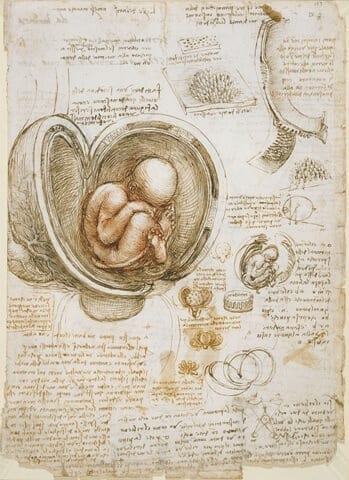
Leonardo Da Vinci had one of the greatest minds in history. Accomplished in so many fields of both the arts and science, he challenged contemporary thinking, and was one of the early Renaissance artists to use dissection of corpses in order to understand the human form. His anatomical drawings reveal a sublime talent at drawing what he saw and showing how the human body was assembled. He demonstrated the framework of bones held together by ligaments, the layers of muscle that connected to tendons to make the joints move, and the network of nerves and blood vessels. Although he could not determine much more about physiology than joint mechanics (he was also an engineer), his drawings show a deep understanding of human anatomy. In this light, however, his depiction of the uterus is astounding. His most famous image of female anatomy is of a pregnant uterus with a mid or late trimester fetus inside (figure). While there is nothing wrong with the depiction of the fetus, there is a problem with the uterus. It is a perfect sphere. Why would Leonardo draw the uterus as a perfect sphere?
To understand the answer we need to reflect on thinking at the time Leonardo was dissecting. In the 1500s when Leonardo was dissecting cadavers the Renaissance was underway, but it was an artistic revival and challenge to previous artistic techniques and subject matter. At the time the Church in Europe dominated thinking, relying on old dogma and vigorously resisting change. The Earth was the center of the Universe and Man the center of the world.
Dissection at the time was proscribed in many places and undertaken by artists at great peril to their safety from authorities. Some universities were undertaking dissection but using the writings of the Roman physician Galen to find their way about. These dissections seemed less an exercise of inquiry and more an affirmation of existing knowledge. In these dissections the appendix regularly caused consternation, because it was not described by Galen who dissected pigs. Galen was right. The cadaver was wrong! Given that the cadavers were commonly bodies that no one wanted to pay for burial, from criminals and vagrants, the interpretation was that they were deviants and therefore not perfect anatomically. Despite the evidence in front of them the learned men of the day denied the existence of the appendix.1
It is in Leonardo’s era of Church dominated thinking and tightly held dogma, despite observational evidence to the contrary, that Leonardo drew his spherical uterus. The uterus was the spiritual center of Woman and so it had to be a perfect sphere. Even one of the greatest thinkers of all time could not let go of contemporary dogma and draw what he saw.
What are the implications of accepting dogma for medicine in 2017? There is no doubt that the establishment of scientific methods and the rise of evidence-based practice has supplanted the need for much of the earlier dogma. However, there is still much we don’t know and plenty of fragments for which we are good at filing in the gaps, without necessarily being sure. As a medical student in the 1980s we were taught the accepted dogma that acid=(peptic) ulcers and (peptic) ulcers=acid.2,3 Interestingly Dr John Lykoudis a Greek physician, treated over 30,000 patients with peptic ulcer disease using antibiotics, but was considered a rogue practitioner and vilified by his learned colleagues.4 Only later were we shown through scientific method that bacteria were the main player and acid only partly responsible for peptic ulcer disease.5 As pediatric trainees in the 1990s we withheld food from patients with diarrhea. Unthinkable now. In 2017 we might think smugly that we know so much and that our dogmatic teachings are based on fact. But are they? Consider your own field and the teachings and disease pathways. How much of what we consider correct will be shown to be wrong in later years. Even the greats, like Leonardo are susceptible. Be careful your preconceived ideas and dog(ma) does not come back to bite you!
References
- Cambridge Illustraed History of Medicne. Cambridge, United Kingdom: Cambridge University Press, 2001.
- Piper DW, Fenton BH. Antacid therapy of peptic ulcer. ii. an evaluation of antacids in vitro. Gut 1964;5:585-9.
- Piper DW, Stiel MC, Builder JE. The electrophoretic pattern of normal human gastric juice and of the gastric juice of patients with gastric ulcer and gastric cancer. Gut 1963;4:236-42.
- Rigas B, Feretis C, Papavassiliou ED. John Lykoudis: an unappreciated discoverer of the cause and treatment of peptic ulcer disease. Lancet 1999;354(9190):1634-5.
- Marshall BJ, Warren JR. Unidentified curved bacilli in the stomach of patients with gastritis and peptic ulceration. Lancet 1984;1(8390):1311-5.
JOHN MASSIE, A/Prof, is a paediatric respiratory physician at the Royal Children’s Hospital (RCH), Melbourne and deputy chair of the RCH clinical ethics committee. John has published extensively in the field of paediatric respiratory medicine, cystic fibrosis, and ethical issues relating to paediatric respiratory medicine. John is a Clinical Associate Professor at the University of Melbourne and Research Fellow at the Murdoch Children’s Research Institute.

Leave a Reply