
Grace O’Connor
Richard Brown
Halifax, Nova Scotia, Canada
Introduction
From: Meier, E. “The cartographers of dead thought.” Max Planck Research, 2 (2014): 70-1.
Oskar and Cécile Vogt were pioneering neuroscientists who established a brain research center in Berlin, Germany, in the early twentieth century. Their work advanced the field of neuroscience through studies on brain architecture and function, including the mapping of architectonic fields and the study of brain disease pathologies.1 Cécile Vogt’s research earned her recognition as an early female scientist. The Vogts’ daughters, Marthe and Marguerite, likewise pursued successful careers in science. Marthe did extensive work on the function of acetylcholine in the nervous system and the function of the kidney in blood pressure regulation.2 Marguerite advanced Drosophila research in genetic inheritance. She later focused on polio virology alongside Renato Dulbecco, ultimately facilitating the polio vaccine and the development of cancerous cell lines.2 The Vogt family’s contributions to neuroscience remain profoundly influential, yet are often overlooked in historical discourse.
Oskar and Cécile
Oskar Vogt (1870–1959) was born in Husum, Schleswig, and attended the University of Jena to study evolutionary biology and comparative anatomy under Ernest Haeckel (1834–1919).1,3 In 1893, he completed his medical dissertation on the corpus callosum under the supervision of Otto Binswanger (1852–1929) and in 1894, he moved to Zurich to study under August Forel (1848–1931).1 Forel was interested in the use of hypnotherapy for mental illness, leading Vogt to study the relationship between hypnotism and brain anatomy. He would become known for his work on introspective hypnotism.1,4 Forel was Vogt’s greatest mentor, sharing similar research interests and career trajectories. Vogt then spent a short time studying brain myelinogenesis in Leipzig with Paul Flechsig (1847–1929), after which he worked during the summer of 1896 as a physician in Alexandersbad, one of the many fashionable spas in Germany.1 It was here that he met FA Krupp (1854–1902), a member of the wealthy Krupp family who owned many mines, arms factories, and railways throughout Germany. Oskar formed close ties with several members of the family and served as their personal physician.1
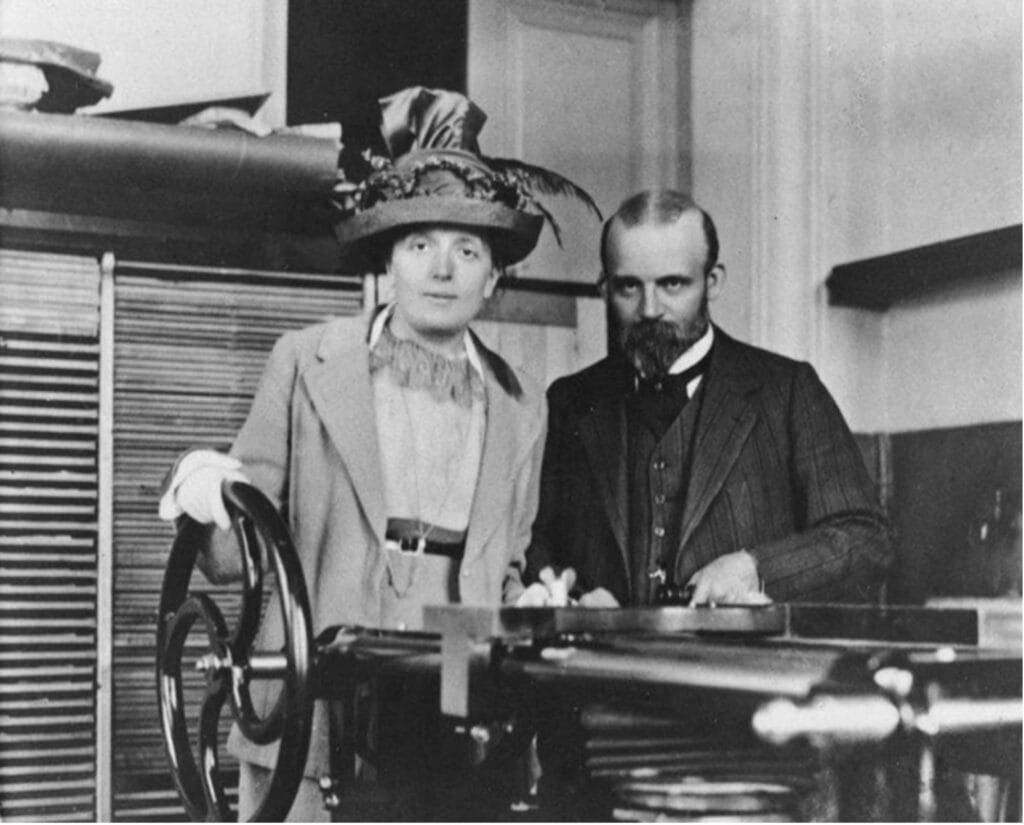
In 1896, Vogt moved to Paris to continue his work on brain structure and hypnosis therapy in the laboratory of Joseph Jules Dejerine (1849–1917). During this time, he met his future wife and lifelong academic collaborator, Cécile Mugnier (1875–1962), born in Annecy, France.3 She earned her baccalaureate degree in science and was one of the first women to enter the Faculté de Médecine in Paris. She completed her dissertation on the myelination of the cerebral hemispheres, supervised by Pierre Marie (1853–1940). Cécile and Oskar met in Paris in 1898 and, despite their language barrier, were married in 1899.1,3
Establishing the research lab
In 1899, the Vogts’ apartment in Magdeburger Strasse, Berlin served as both private practice for Oskar’s hypnotism work and a shared research laboratory called the Neurological Central Station (Neurologische Zentralstation), which focused on brain anatomy and physiology (Figure 1).1 In 1902, the station was incorporated into the physiology department at the University of Berlin with funding from the Krupp family. By 1914, the Vogts’ laboratory had expanded into the Kaiser Wilhelm Institute (KWI) for Brain Research (Kaiser-Wilhelm-Institut für Hirnforschung) in Berlin.1

Architectonics
Using measures such as fiber density and myelin sheath thickness to define myeloarchitectonic fields, the Vogts provided a comprehensive view of brain myelination.3 They further analyzed the arrangement, number, size, and form of nerve cells within the laminae of the cortex (cytoarchitecture) and the corresponding relationships of the myelinated nerve fibers (myeloarchitecture). Using their criteria for cytoarchitecture and myeloarchitecture, they were able to map the brain into structurally distinct regions, known as architectonic fields, and determine the corresponding physiological mechanisms (Figure 2).3 To further refine their architectonic maps, the Vogts conducted electrophysiological mapping in both humans and monkeys to quantify over 200 architectonically distinct areas.5,6
Korbinian Brodmann and the Vogts
Korbinian Brodmann (1868–1918) joined the Vogts’ research faculty in 1901 to study cytoarchitecture. Brodmann published the now-famous concept of Brodmann’s areas in 1909, which defined forty-three areas of the human cortex.7 Brodmann’s simplified numerical cortical map and its widespread use during World War 1 as an orientation tool for military neurosurgeons made his work more accessible and easier to apply in research and clinical applications.7,8
Lenin’s brain
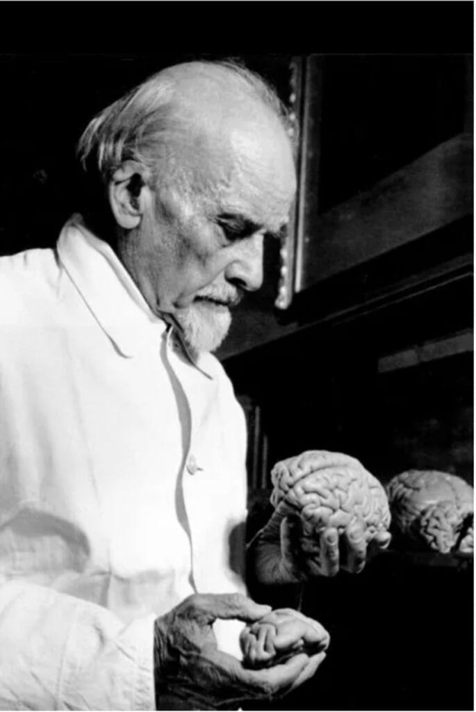
After the death of Vladamir Ilyich Lenin (1870–1924), Soviet officials, believing that his genius must have had an anatomical underpinning, invited Oskar Vogt to examine his brain in detail (Figure 3).1 Vogt used cytoarchitectonic and myeloarchitectonic techniques to determine that Lenin’s brain had large pyramidal neurons in the third cortical layer and noted that there was a particular increase in richness and distinctive configurations of the sulci and gyri within the frontal lobe.1 The story of Lenin’s brain has been given a fictional account by Spengler (1991).9
Dealing with the Nazis
Oskar Vogt faced significant backlash for his sympathies and collaboration with Russia regarding Lenin’s brain as well as his general leftist ideologies. In 1934, Oskar was discharged from his position as director of the Kaiser Wilhelm Institute, an order signed by Adolf Hitler (1889–1945).10 Despite this setback, the Vogts’ passion for research endured. With financial assistance from the Krupp family, they built a new institute referred to as the Vogt Institute in Neustadt, Germany.1 The Vogts continued their research on architectonics in their new brain institute, publishing research on the aging of nerve cells from factors relating to arteriosclerosis.11
Cécile Vogt’s crucial contributions
While Oskar Vogt often garnered much of the credit for their lab’s accomplishments, Cécile supervised the anatomy and neurochemistry departments of the institute as well as its finances.1 Importantly, she identified forty nuclei of the thalamus in primates12 and revolutionized the treatment of psychiatric disorders through her concept of the use of pharmacotherapeutics. Although hysteria was initially characterized as a gender-related issue, Cécile redefined it as a somatic problem involving the function of the corpus callosum that could be treated with pharmacotherapy.3
Marthe Vogt
The Vogts’ first daughter, Marthe (1903–2003), earned degrees from the University of Berlin in medicine (1928) and chemistry (1929).2 In 1931, she joined the chemistry department of the Kaiser Wilhelm Institute, working alongside her parents. At the KWI, she investigated the effects of drugs on the central nervous system.13 She then moved to England where she worked at the National Institute for Medical Research alongside Henry Dale (1875–1968) and Wilhelm Feldberg (1900–1993), where they demonstrated that nerves in the spinal cord elicited muscle contraction through the release of acetylcholine.14 Later at Cambridge, Marthe worked with EB Verney on uremia, demonstrating that a substance later identified as renin was released from the kidney and led to the condition.15 In the laboratories of the Pharmaceutical Society of Great Britain, Marthe’s work focused on the effects of the adrenal gland, and she demonstrated the effects of adrenocorticotropic hormone in response to stress.11 In 1946, her innovative catecholamine assays revealed the heterogeneous distribution of epinephrine and norepinephrine in the brain.13 She concluded that the distribution of norepinephrine could not be accredited to sympathetic vasomotor nerves.
Marguerite Vogt
Marguerite, the younger Vogt daughter, was born in 1913. When she was only fourteen, she began describing mutations among Drosophila, or fruit flies.2 She earned her medical degree from the University of Berlin in 1924, and then worked at the KWI studying the structure and function of the ring gland and genetic mutations in Drosophila, even developing her own morphogenetic assay.16,2 In the early 1950s, she began to work with Renato Dulbecco in California, and they became the first to observe the formation of plaques in tissue cultures as a result of the polio virus, allowing them to grow polio in vitro.17 Dulbecco and Vogt also worked on polyomavirus, a family of DNA viruses, which revolutionized cancer research through the ability to transform healthy cells into cancerous ones without killing the line.18 The two would collaborate on the work that eventually earned Dulbecco a Nobel prize in 1975, by demonstrating the transfer of genetic material among tumor viruses.
Importance of the Vogts’ research today
The four members of the Vogt family made significant contributions to neuroscience. Oskar and Cécile expanded the study of brain architecture, function, and disease pathologies. Their work on architectonics allowed for a greater understanding of anatomical areas and projection fields, earning them Nobel Prize nominations in 1922 and 1926.19,20 Marthe’s work on acetylcholine and assay development led to the discovery of epinephrine and norepinephrine. Marguerite made significant contributions to the study of Drosophila genetics, discovered the plaque formations in polio viruses, and demonstrated cancerous transmission between cells. The importance of their work exceeds their individual discoveries as well as exemplifies the advances of women in science (Figure 4).
References
- Klatzo I. “Cécile and Oskar Vogt: The visionaries of modern neuroscience.” Acta Neurochirurgia Supplement 80, (2002): 1-130.
- Rubin, RP. “The Vogt family: Creators of diverse paths for women in biological research.” Journal of Medical Biography 25, (2017): 252-260. 10.1177/0967772017731300
- Haymaker, W. “Cécile and Oskar Vogt, On the Occasion of her 75th and his 80th birthday.” Neurology 1, (1951):179–204. https://doi.org/10.1212/wnl.1.5.179
- Mayer, Andreas. Sites of the Unconscious: Hypnosis and the Emergence of the Psychoanalytic Setting. (University of Chicago Press, 2013), 182.
- Vogt, C., and Vogt, O. “Allgemeinere Ergebnisse unserer Hirnforschung.” Journal für Psychologie und Neurologie 25, (1919): 292-398.
- Vogt, C., and Vogt, O. “Die vergleichend-architektonische und die vergleichend-reizphysiologische Felderung der Großhirnrinde unter besonderer Berücksichtigung der menschlichen.” Die Naturwissenschaften 14, (1926): 1190-4.
- Mueller, T., and Kanis-Seyfried, U. “On the life and work of Korbinian Brodmann (1868–1918).” Journal of the History of the Neurosciences 28, (2019): 307-18. https://doi.org/10.1080/0964704X.2019.1589689
- Olry, R. “Korbinian Brodmann (1868–1918).” Journal of Neurology 257, (2010): 2112-3. https://doi.org/10.1007/s00415-010-5702-x
- Spengler, T. Lenin’s Brain. Translated by Shaun Whiteside. London: Hamish Hamilton, 1991.
- Stahnisch, FW. “Morphological Research Directions at Neuroscience-Related Institutes of the German Max Planck Society, 1948–2002.” Anatomia 3, (2024): 301-32. https://doi.org/10.3390/anatomia3040024
- Vogt, C, Vogt, O. “Ageing of Nerve Cells.” Nature 158, (1946): 304. https://doi.org/10.1038/158304a0
- Vogt C. “La myéloarchitecture du thalamus du cercopithèque.” Journal de Psychologie Neurologiques 12, (1909): 285-324.
- Cuthbert, AW. “Marthe Louise Vogt 8 September 1903 – 9 September 2003.” Biographical Memoires of the Fellows of the Royal Society 51, (2005): 409-23. https://doi.org/10.1098/rsbm.2005.0027
- Dale, HH, Feldberg, W, Vogt, M. “Release of Acetylcholine at Voluntary Motor Nerve Endings.” The Journal of Physiology 86, (1936): 353-80. https://doi.org/10.1113/jphysiol.1936.sp003371
- Verney, EB and Vogt, M. “An experimental investigation into hypertension of renal origin, with some observations on convulsive ‘Uraemia.’” Quarterly Journal of Experimental Physiology and Cognate Medical Sciences 28, (1938): 253-303.
- Vogt, M. “Bemerkung zum Corpus allatum von Drosophila.” Die Naturwissenschaften 28, (1941): 725-726.
- Dulbecco, R, and Vogt, M. “Plaque formation and isolation of pure lines with poliomyelitis viruses.” Journal of Experimental Medicine 99, (1953): 167-82. https://doi.org/10.1084/jem.99.2.167
- Oransky, I. “Marguerite M. Vogt.” The Lancet, 370 (2007): 1122. 10.1016/S0140-6736(07)61500-1.
- The Nobel Prize. “Nomination Archive,” 1922. https://www.nobelprize.org/nomination/archive/show.php?id=8901.
- The Nobel Prize. “Nomination Archive,” 1926. https://www.nobelprize.org/nomination/archive/show.php?id=6479.
GRACE O’CONNOR is completing her honors BSc in neuroscience at Dalhousie University. Her research interests involve fMRI studies of addictive processes.
RICHARD E. BROWN is a professor of psychology and neuroscience at Dalhousie University and conducts research on the neurobiology of behaviour. His research is on mouse models of neurodegenerative disorders. He is also interested in the history of neuroscience.