
Saty Satya-Murti
Santa Maria, California, United States
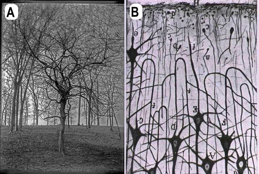
B. Neuronal forest: Cajal’s 1923 Golgi stained image (inverted) of pyramidal cells of rat cerebral cortex. National Library of Medicine.
Plants have long inspired humans. Early natural scientists were struck by similarities between structural anatomy in animals and the botanical arrangements of plants. Bent tree trunks, spreading foliage, forested canopies, and curvaceous tendrils inspired them to draw artistic comparisons, both obvious and imagined, between plants and animals. Phenomenologists coined mimetic labels for some human conditions based on what they had observed in plants. Some terms were just linguistic coincidences, but many others had common, logical roots.
Cajal’s neuronal forests
In the late nineteenth and early twentieth centuries, Camillo Golgi’s (1843–1926) silver stain of mammalian brain sections provided stunning black-and-white silhouettes of cells in the cerebral cortex and cerebellum. Using a Golgi stain, Santiago Ramon y Cajal (1852–1934) described the extensive repeated branching of neurons, axons, and dendrites. This prompted him to write exuberantly about the “neuronal forest” populated with “gleaming needles, [and] filaments that were smooth and thin or thorny and thick,” and branches covered with “spine[s].”1,2 Cajal excelled at artistic representations of reality.3 His poetic vocabulary effectively expressed both his love of nature and his passion for histologic precision. Indeed, a veritable “neuronal forest” existed in mammalian brains. (Figure 1) Early morphologists used the term “arbor vitae” to denote the extensive arborization patterns seen in microscopic sections of the nervous system. Arbor vitae cerebelli, a tree-like fractal branching of cerebellar white matter, draws scientific fascination even today.4 Both Golgi and Cajal received the Nobel Prize in Physiology and Medicine in 1906.
Andry’s orthopedic tree
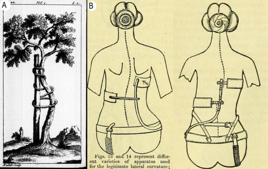
B. An early example of bracing “apparatus” used for scoliosis correction. From Taylor, Charles Fayette and Royal College of Surgeons of England. The Pathology and Treatment of Lateral Curvature of the Spine (New York: New York Print. Co., 1868). Via the Internet Archive.
In the mid-1700s, French physician Nicholas Andry (1658–1742) observed how a crooked young tree trunk had been splinted to reduce its curvature and aid its vertical growth. The idea of splinting children’s limbs or spine for “preventing and correcting the deformities” could have arisen based on such observations. Andry’s mimetic creativity led him to coin the term “orthopedics” (orthos = straight, paidios = a child).5,6 Awareness of “crippling deformities” in the young preceded the coinage of this term.. Andry’s observations may not have been the earliest to realize the therapeutic potential of splinting; however, his 1741 classic book used the term “Orthopedie” for the first time. This book has an illustration of his “orthopedic tree,” whose metaphoric implication has since come to symbolize the modern practice of orthopedics. (Figure 2) One example is the treatment of scoliosis with braces, casts, instrumentation, and exercise. The goal is to halt the progression of curvature of the spine, which may have originated from the observation of splinted trees.
Bumps on barks and crusts on cutis
“Freddie Fungus and Alice Algae took a lichen to each other.”
—Mnemonic used by naturalists as a teaching tool
Tree bark and human skin, both highly visible integuments, became a favored domain for botanical analogies. Both human and plant integuments exhibit evidence of fungal/mycotic presence. Two terms, lichen and mycosis, merit specific emphasis. Plant lichen is a generally harmless, crustaceous growth on tree bark. Descriptions of plant lichens were first recorded around 1562. In people, lichen is a term for chronic autoimmune conditions of the skin, nails and mucosa. Descriptions of this condition were first noted around 1798, and the Oxford English Dictionary refers to lichen simplex as a “skin disease characterized by patchy areas of thickening of the epidermis, typically occurring in response to repeated rubbing or scratching.”7
Early observers apparently thought that lichens, human or plant, looked as if something “seemed to lick the affected organism,” the affected part being the bark or the skin.8 The Greek noun “leikhen” means “to lick.” Even in 1818, Robert Willan’s cutaneous disorders treatise had recognized “the great irritability of the skin” in lichen, “manifest from the facility with which the papulae are enlarged into temporary wheals by strong friction, which the itching and tingling compel the patient to resort to.” This response to rubbing or scratching may have been an earlier description of Koebner’s phenomenon that predated Heinrich Koebner’s (1838–1904) eponymous description in 1876. However, the external appearance of “lichen” in plants and humans was the sole basis for the linguistic coincidence. There is no pathobiological or etiological connection between them. (Figure 3) In the natural world, lichens may grow on any surface, dead or living, from trees to gravestones to skulls. Lichens that grew on human skulls were part of therapeutic armamentaria in the past; Frederick Chopin was administered skull lichen tea for his congestive heart failure.9,10
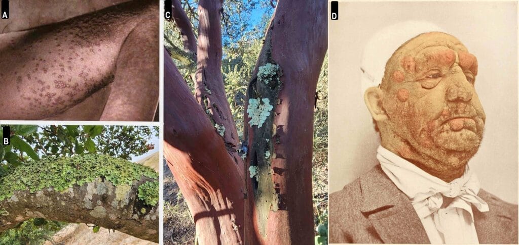
B, C. Plant lichens. From author’s collection.
D. Mycosis fungoides (1905). Via Wikipedia.
The term “myco” derives from the Greek mýkēs, meaning mushroom or fungus. In 1806, the French dermatologist Louis Alibert (1768-1837) noted “small tubercles … with smooth glazed surface …[of] a light brownish color” in in a fifty-six-year-old Parisian patient. The tubercles seemed to “multiply to such a point that we can count fourteen in the face.” To Alibert, they resembled mushrooms, and so he termed the condition “mycosis fungoides.”11 We know now that the similarity to mushrooms stops with external appearance because mycosis fungoides, notwithstanding its namesake, is a cutaneous T-cell lymphoma. Much like Cajal, Alibert was also a tree lover. He devised a classification system for skin diseases in the mode of Linnaean taxonomy and called it the “Tree of Dermatosis.”
Hives (urticaria) affect almost 20% of humans at some point in their lives. There are multiple etiologies, but one cause is exposure to the stinging nettle (Urtica dioica), a plant with fine, hollow-bore hairs on its serrated leaves and stems. The term “urtica,” derived from Latin, means “to burn.” Skin contact with this edible, common, perennial plant causes irritation and itching due to release of histamine and other inflammatory mediators.12 Human urticaria owes its appellation to what was observed after exposure to the plant Urtica dioica.
Besides the brain, bones, and skin, other examples of morphologic similarities between plant structures and human anatomy have led to shared labels. The renal calyx reminded observers of the calyx of developing flowers, and a congested liver was reminiscent of a nutmeg. In 1882, Scottish bacteriologist Alexander Ogston (1844–1929) examined pus from surgical wounds and found organisms whose growth and clustering resembled bunches of grapes under the microscope. He named them Staphylococcus; staphyle in Greek meaning “bunch of grapes.”
Human gall and tree gallare exceptions to the logic behind mimetic terminology. Tree galls, small pebble-sized excrescences on branches formed by maturing wasp eggs, bear no connection except a linguistic coincidence with human gall (bile).
Anthropic analogies to plants beyond visual resemblance are recognizable in other ailments such as maple syrup urine disease (MSUD) and pediatric-age fractures. The urine of a person with MSUD has a distinct odor, similar to that of maple sugar, California cudweed, and fenugreek. Greenstick fractures occurring in children resemble the one-sided rupture of tender plant trunks. They do not sever the entire integrity of the bone or the plant.
Our curious and observant forebears have thus given us a rich medical vocabulary that owes its origins, in part, to the diverse visual beauty of the plant world.
References
- DeFelipe, Javier. “Cajal and the Discovery of a New Artistic World.” In Progress in Brain Research, 203:201–20. Elsevier, 2013. https://doi.org/10.1016/B978-0-444-62730-8.00008-6.
- Garrido, Eduardo. “Cajal and His Love for Nature: A Sentimental Essence in the Legacy of Neurosciences.” Frontiers in Neuroanatomy 18 (July 18, 2024): 1408783. https://doi.org/10.3389/fnana.2024.1408783.
- Reilly, Cate. “Neuromimesis: Picturing the Humanities Picturing the Brain.” Frontiers in Integrative Neuroscience 16 (October 14, 2022): 760785. https://doi.org/10.3389/fnint.2022.760785.
- Maryenko, Nataliia. “Arbor Vitae Cerebelli: Fractal Properties and Their Quantitative Assessment by Novel ‘Contour Scaling’ Fractal Analysis Method (an Anatomical Study).” Translational Research in Anatomy 37 (November 1, 2024): 100352. https://doi.org/10.1016/j.tria.2024.100352.
- Ponseti, Ignacio V. “History of Orthopaedic Surgery.” The Iowa Orthopaedic Journal 11 (1991): 59–64.
- Fadlurrahman, Manaf. “Homini Verminoso, Who Created ‘Orthopaedia.’” Hektoen International, May 7, 2019. https://hekint.org/2019/05/07/homini-verminoso-who-created-orthopaedia/.
- Oxford English Dictionary, s.v. “lichen simplex (n.),” June 2024, https://doi.org/10.1093/OED/6788540576.
- Oxford English Dictionary, s.v. “lichen (n.), Etymology,” December 2024, https://doi.org/10.1093/OED/2201141136.
- Modenesi, P. “Skull Lichens: A Curious Chapter in the History of Phytotherapy.” Fitoterapia 80, no. 3 (April 2009): 145–48. https://doi.org/10.1016/j.fitote.2008.12.005.
- Arnold, Wilfred. “Chopin’s Heart.” Hektoen Innternational, Spring 2011. https://hekint.org/2017/01/30/chopins-heart/.
- Karamanou, Marianna, Theodora Psaltopoulou, Gregory Tsoucalas, and George Androutsos. “Baron Jean-Louis Alibert (1768-1837) and the First Description of Mycosis Fungoides.” J Buon 19, no. 2 (2014): 585–88.
- Grauso, Laura, Bruna De Falco, Virginia Lanzotti, and Riccardo Motti. “Stinging Nettle, Urtica Dioica L.: Botanical, Phytochemical and Pharmacological Overview.” Phytochemistry Reviews 19, no. 6 (December 2020): 1341–77. https://doi.org/10.1007/s11101-020-09680-x.
Acknowledgement
I thank Susan Tuttle, California Naturalist and Certified Interpretive Guide at The Los Flores Ranch Park, City of Santa Maria, for her patience in educating me about the natural beauty and features of California’s Central Coast.
SATY SATYA-MURTI, MD, FAAN, is a clinical neurologist and health policy consultant. Following retirement, Saty has spent time researching cognitive biases, the social underpinnings of clinical medicine, Progressive Era medicine, and forensic sciences. He enjoys grandparenting, solar cooking, and volunteering.
One response
Saty, thank you for sharing! This is so interesting. Another reminder that all living things are truly amazing.