
US CDC Public Health Image Library.
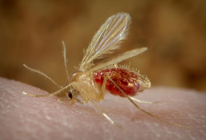
Sand flies are about three millimeters long and gray, brownish or golden, with long, piercing mouthparts adapted for sucking blood from their hosts. These seemingly innocuous creatures, classified as belonging to the genus Phlebotomus or Lutzomyia, have long legs and hairy wings held in a vertical V-shape when at rest. They seek moist resting places such as caves or tree holes, and feed mainly at night. After a blood meal, the female sand fly can lay thirty to seventy eggs that hatch into larval and pupal stages in one or two weeks. Adult flies can transmit some twenty species of Leishmania protozoa capable of causing illness.1 Mainly considered a disease of poverty and poor sanitation, in Asia, Africa, South America, and Southern Europe, leishmaniasis has also affected military personnel and tourists as well as refugees. According to estimates from the World Health Organization, it has infected some four to twelve million people in some 98 countries.1,2
The parasites cause three kinds of leishmaniasis illnesses: 1. A disease of the skin occurring in over one million persons per year1 and consisting of either a single sore that heals slowly, leaving an ugly scar, or of widespread lesions that resemble leprosy; 2. A mucocutaneous form of leishmaniasis in which the nose and mouth bear the brunt of the disease; 3. Visceral leishmaniasis or kala-azar, severe and often fatal, characterized by fever, anemia, enlargement of the liver and particularly of the spleen, which may become huge.1,2
Fossil evidence suggests that the Leishmania parasites emerged during the Mesozoic era over 250 million years ago.2 They caused disease in antiquity, as shown on tablets from King Ashurbanipal of Assyria in the seventh century BC.2 Widespread also in the Middle Ages, the disease was called by Arab scholars “Aleppo boil” or “Baghdad boil,” names that persist even today. This long historical impact of the disease commands respect. Alexander Russell, after examining a Turkish patient, gave a clinical description in 1756, followed in 1824 by Dr. William Twining in Bengal. Indians called it “Assam fever” after it spread to that particular region, and then kala-azar or black sickness, reflecting the characteristic darkening of the skin in lighter-skinned patients. At first, kala-azar was mistaken for malaria, but late nineteenth-century doctors recognized its distinct symptoms—fever, weight loss, and swelling of the spleen and liver—as well as its devastating effects on millions of people across the globe, particularly in tropical and subtropical regions.
David Douglas Cunningham, a scientist in Britain (1885), and Peter Borovsky, a Russian military surgeon working in Tashkent (1898), appear to have been the first to have seen the parasite under a microscope, but credit is traditionally given to William Leishman and Charles Donovan (1903), hence the name Leishmania donovani.2 Leishman was a Scotsman who served with the Royal Army Medical Corps in India and did research on enteric fever and kala-azar. Returning to the United Kingdom in 1897, he became an Assistant Professor of Pathology at the Army Medical School, where he invented the simplified method of staining blood for parasites with a combination of methylene blue and eosin that became known as the Leishman stain. In 1901, while examining pathologic specimens of a spleen from a patient who had died of kala azar, he observed the oval bodies causing the disease. As these were also described by Charles Donovan of the Indian Medical Service, they are now known as Leishman–Donovan bodies.
The early twentieth century saw devastating epidemics of the disease, especially in India, with mortality rates as high as 95%. These outbreaks led to increased research efforts to understand and combat the disease. In 1915, Upendranath Brahmachari developed the first effective treatment using antimony compounds.2 Later, amphotericin B and pentamidine became available as alternative drugs. World War II brought renewed attention to kala-azar as soldiers deployed to endemic areas were infected. Currently, visceral leishmaniasis is most often caused by L. donovani and L. chagasi, but also by several other varieties. Clinical features vary somewhat in different groups, and several additional drugs are being tried to control the disease.1,2
In the latter half of the twentieth century, the World Health Organization launched initiatives to combat the disease, but the rise of drug resistance, environmental changes, and population displacement contributed to ongoing challenges. However, the relentless efforts of researchers and medical professionals have not waned. Research now focuses on developing more effective treatments, improving diagnostic techniques, creating a vaccine, and reducing the threat of sand flies, which, though small in size, constitute a colossal threat to humankind. This ongoing research instills hope for a future free from the burden of leishmaniasis.
References
- Pearson RD and de Queiroz Sousa A. Clinical Spectrum of Leishmaniasis. Clinical Infectious Diseases Jan 1996;22 (1):1-11.
- Leishmaniasis. Wikipedia. https://en.wikipedia.org/wiki/Leishmaniasis
Leave a Reply