
JMS Pearce
Hull, England
Likened to the small intestines, in ancient times the gyri of the brain were named “coils” by Greek physicians and anatomists. Vesalius in the sixteenth century amplified the description in the celebrated De humani corporis fabrica. Thomas Willis in Cerebri anatome (1664) radically changed the accepted view that cognitive and somatic functions were seated in the cerebral ventricles when he stated “movement is initiated in the cerebrum.”1
Gratiolet,2 Vicq d’Azyr,3 and later Magendie4 were amongst the first to describe the convolutions, noting the differences in morphology in different animals. In France, before Paul Broca’s studies on the localization of language, Jean-Baptiste Bouillaud (1796–1881)5 and his son-in-law Auburtin located language in the frontal lobes. They acknowledged the import of the works of Franz Gall6 and Johann Spurzheim.
The fissure of Rolando7

The attachment of eponyms to cerebral fissures had started with the lateral fissure of Sylvius in 1663, though this had been described by Caspar Bartholin (1585–1629), a Danish anatomist. The second structure to be so dignified was the fissure of Rolando in 1839.8 Luigi Rolando (1773–1831) observed the precentral and postcentral gyri on either side of the great central fissure. (Fig 1)
Following his teacher, the versatile Vincenzo Malacarne (1744–1816), Rolando referred to the convolutions as enteroid processes, a term not very different from that of Erasistratus (304–250 BC).
Grays Anatomy states that the central sulcus (fissure of Rolando) (Fig 1) is situated about the middle of the lateral surface of the hemisphere. It runs sinuously downward and forward, and ends a little above the posterior ramus of the lateral Sylvian fissure. It forms an angle of about 70° with the median plane.
At the turn of the nineteenth century the gross structure of the brain was emerging, but its functioning lay in a crucible of ignorance. Rolando, impressed by Galvani’s demonstration of electricity in the production of movement in experimental animals, deduced that since the brain generated movement, it might generate electricity. He experimented on pigs, guinea pigs, goats and sheep. In 1809, he applied an electric current through a wire placed in parts of the brain; this caused violent limb contractions, then stupor. Because he realized the experimental damage inflicted by his procedure, he was at first cautious in his conclusions, but he deduced that nervous structures are connected in a network of nervous fibers that functioned by electrical impulses. He had however, shown that movement was primarily generated by the precentral cortex adjacent to the central sulcus.9
Rolando’s experiments were largely unknown until Magendie published a French translation in 1823, reprinted by Flourens. Rolando had correctly located motor activity to the precentral cortex. It fell to Francois Leuret to give due credit to Rolando:
Between these two convolutions exists a furrow that separates them… it is as constant as the Sylvian fissure. I have called this furrow the fissure of Rolando, because it was this anatomist who first described it in man, in whom it is still more developed than in the monkey.10,11
Subsequently, motor functions were differentially apportioned to the primary motor cortex, premotor cortex, and supplementary motor area. The conflicting notions of the motor cortex as the “organ representing movements” rather than precisely demarcated muscle activities were later clarified—mainly by Hughlings Jackson and David Ferrier in the nineteenth century.
Cerebellum
Many of Rolando’s experiments were on the cerebellum, basal nuclei, and midbrain. In the early nineteenth century, the function of the cerebellum was unknown until Rolando’s investigations.12 He noticed the resemblance of the lamellae of the cerebellum to Voltaic piles, and speculated that a nervous fluid originated and then was led “through the different nerves and brought to stimulate the muscles subservient to locomotion.” If the cerebellum generated electricity for muscle activity, its removal might produce paralysis. He therefore removed the cerebellum in a goat and observed that it could no longer stand up: “non altrimenti che se fosse paralitico” (not other than if it was paralyzed). Rolando observed that minor damage of the cerebellum caused staggering, but destruction caused total locomotor paralysis in his animals.11 He therefore believed the cerebellum determined not only the regulation of, but also the strength of movement. Although an inaccurate view, this was a step forward from the prevailing notions of the cerebellum as controller of intellect, sensation, or vital forces.
Meanwhile, acting on orders of Napoleon Bonaparte to test Franz Gall and Johann Spurzheim’s contentious theories of cerebral localization,7,8 Marie-Jean-Pierre Flourens removed the cerebellum in animals; he observed that movement was retained but equilibrium and co-ordination were impaired.13 In a series of somewhat crude ablation experiments, Flourens criticized Rolando’s conclusions. The physiology of cerebellar motor and cognitive functions was later refined by studies in man by Gordon Holmes,14 JC Eccles, and Jeremy Schmahmann.
Flourens also removed the cerebrum in pigeons, showing maintenance of reflexes with loss of cerebration. He opposed the idea of cerebral localization, believing that the brain functioned as a whole. Thus arose the concept of “cerebral equipotentiality”: an early holistic hypothesis of brain function.11
Substantia gelatinosa
Another important advance was his account of the substantia gelatinosa Rolandii in the dorsal horns of the spinal cord:
A particular grey matter in the posterior third of the posterior horns, which he noticed was, more gelatinous…of a different colour…15
The substantia gelatinosa is a fine network of interneurons that relate to pain and temperature sensation and is rich in substance P and opiate receptors. It was subsequently named after Rolando.11
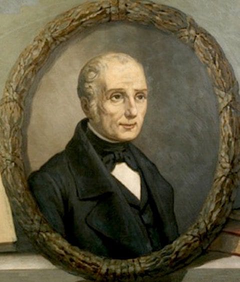
Luigi Rolando
Luigi Rolando (Fig 2) was born in Turin. After completing his medical studies he concentrated on experimental anatomy.11 He studied in Florence and became physician to the King of Savoy in Turin. The invasion of Italy by Napoleon drove him into exile in Sardinia, where in 1804 he became professor of theoricopractical medicine at Sassari. After the Napoleonic wars, he returned to Turin in 1814 as professor of anatomy where he is commemorated by the Luigi Rolando Museum of Human Anatomy.16
Recognition did not come easily, since the almost contemporaneous works of Gall and Spurzheim on cerebral localization (and phrenology) and of Flourens on cerebellar function challenged his discoveries.
Rolando died on 20 April 1831, of cancer of the pylorus.
Several neuroanatomical structures bear his name: the Rolandic vein, the Rolandic artery (central sulcal artery), the Rolandic operculum (post-central operculum), the Rolandic area (primary motor cortex), the substantia gelatinosa of Rolando, the fissure of Rolando (central sulcus) and Rolandic epilepsy.
References
- Dewhurst K. Willis’s Oxford Lectures. Oxford: Sandford Publications, 1980.
- Gratiolet P. Memoire sur le pliés cérébreaux de l’homme et des primates. Paris: Bertrand, 1854.
- Vicq d’Azyr F. Traité d’anatomie et de physiologie—avec des planches colores représentant au naturel les divers organes de ‘Homme et des Animaux. F.A. Didot, Paris, 1786.
- Magendie FJ. An elementary compendium of physiology: For the use of students. Trans: E. Milligan. Philadelphia: James Webster, 1824, 104.
- Bouillaud JB. Recherches cliniques propres à démontrer que la perte de la parole correspond à la lésion des lobules antérieurs du cerveau, et à confirmer l’opinion de M. Gall, sur le siège de l’origine du langage articulé. Arch Gen Med (Paris, 1ère série) 1825; 8:25-45.
- Gall F. J. Craniologie ou dicouvertes nouvelles cancernant le cerveau lecrane et les organes. Paris: Necelle, 1807. See also: Dictionnaire des Sciences Medicales. Paris, 1813;4:447.
- Portions of this and later sections excerpted from Pearce JMS, The fissure of Rolando. First published in J Neurol Neurosurg Psychiatry 1999;67(4):528.
- Schiller F. The Rise of the “Enteroid Processes” in the 19th Century: Some Landmarks in Cerebral Nomenclature. Bulletin of the History of Medicine 1965; 39:326-338.
- Rolando L. Saggio sopra la vera struttura del cervello dell’uomo e degl’animali e sopra le funzioni del sistem nervoso. [Essay on the true structure of the brain of man and animals and on the functions of the nervous system.] Sassari, Italy: Stamperia Privilegiata, 1809.
- Leuret F. Anatomie comparée du système nerveux. Paris: Baillière 1839, 1:397-8.
- Pearce JMS. The fissure of Rolando. J Neurol Neurosurg Psychiatry. 1999;67(4):528.
- Rolando L. Osservazioni sul cervelletto. Mem. R. Accad. Sci. Torino, 1825, 29, 163-88.
- Flourens P. Analyse de la Philosophie Anatomique, ou l’on considère plus particulièrement l’influence qu’aura cet ouvrage sur l’état actuel de la Physiologie et de l’Anatomie. Paris: Béchet, 1819.
- Holmes GM. On the clinical symptoms of cerebellar disease. Croonian Lectures. Lancet 1922;I:1177–82, 1231-7 II:59–65, 111-5.
- Rolando L. Ricerche anatomiche sulla struttura del midollo spinale. [Anatomical structure of the spinal cord.] 1824.
- Belloni F. Essays on the history of Italian neurology. ed Belloni (Ist di Storia della Medicine, Milan), 1963, p. 204. Cited in: Pearce, The fissure of Rolando.
JMS PEARCE is a retired neurologist and author with a particular interest in the history of medicine and science.