
Daniel Gelfman
Indianapolis, Indiana, United States
The use of forensic science to determine the etiology and manner of death has been attempted for millennia. Early autopsies involved inspection of the deceased individual and possibly an internal examination. The performance of autopsies has been greatly influenced by religious and political forces.1 There is a record of the autopsy on Julius Caesar described by the historian Suetonius around 150 CE, which had been performed in 44 BCE by (presumably) his physician, Antistius. Using careful observation, this autopsy revealed that Caesar died due to exsanguination from one of his twenty-three knife wounds, specifically the one that apparently severed his heart and aorta.2 Unfortunately, autopsies using observation and dissection alone often do not yield a satisfactory answer as to the cause and manner of death. Progress in the science of forensics has required enormous advances in technology; in knowledge of anatomy, physiology, and pathophysiology; and in techniques for crime scene examination.
Blood spatter, or bloodshed analysis, is a more recent addition to the field of crime scene analysis. The first recorded study of this subject occurred in 1895 when Dr. Eduard Piotrowski at the University of Krakow, Poland, published a paper entitled, “Concerning the origin, shape, direction, and distribution of the bloodstains following Head Wounds Caused by Blows.” This field was prominently influenced in the 1900s by Drs. Paul Kirk and Herbert MacDonell. As it is based on observations of findings that are potentially influenced by many external factors and thus could have more than one interpretation, the precision of this science is controversial. However, most believe that there is useful information derived from blood spatter analysis.
While observation is vulnerable to biased interpretation, it should be noted that progress in medicine has often originated with careful observation. Quality medical care itself requires thoughtful observation. However, the veracity and usefulness of any observation depends on the absence or minimalization of bias. Artists are by their very nature skillful observers and reveal this in their work. The study of art has been used to teach the skill of careful observation in medical education. Interestingly, there are many examples of artists depicting observable medical anatomic and physiologic findings before they were described and understood by the established medical community.3,4,5
The usual “language” of the artist is different from that of the scientist, as the artist uses pictorial or three-dimensional representations to express ideas and findings, generally without words. Only rarely can an artist who is also a scientist, such as Leonardo di Vinci (1452–1519), depict new ideas in artwork and discuss them as well.
While not usually conventional scientists, artists are often aware of advances in science. An understanding of anatomy, physiology, and science in general can influence their work. It has been postulated that pictorial representations of arterial bleeding in Baroque art was influenced by Galileo Galilei (1564–1642), who discovered the “theory of projectile motion” (1604–1608). Caravaggio (1571–1610) depicted a rectilinear bleeding trajectory in Judith Beheading Holofernes (1599) before Galileo’s theory was known. In contrast, Artemisia Gentileschi (1593–1693), who is purported to have known Galileo, depicted the same topic in 1620, after his theory had been revealed, with an arterial bleeding trajectory that was parabolic.6
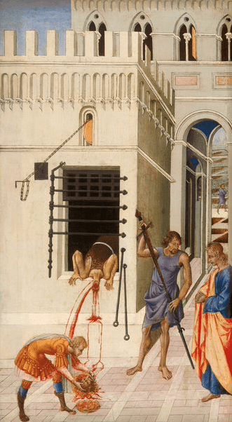
Impressively, 150 years before Galileo’s discovery, a lesser-known Sienese artist of the early Renaissance, Giovanni di Paolo (1398–1482), painted The Beheading of Saint John the Baptist (c. 1455–1460) and revealed a “crime scene” with several anatomic and physiologic phenomena related to bleeding. These include the parabolic trajectory of arterial bleeding as well as what appears to be a specific blood stain pattern from beheading.7 There is evidence that beheading was practiced in neighboring Florence8 and thus it is possible that Giovanni di Paolo witnessed such an event. There is a large, wide stream of blood emanating from the neck of Saint John with a parabolic trajectory, likely from the carotid artery. Next, there is a smaller stream of blood, also in a parabolic trajectory, likely from the smaller vertebral artery. We only see parabolic bleeding from two arterial sources and not the expected four, as these arteries are present bilaterally. A venous bleeding pattern is portrayed in blood that simply flows down from the neck wound. We do not see the source of this bleeding from the neck. All of this is consistent with observation that occurred at a distance without close examination of a human body. There is also significant detail from the blood on the ground to suggest a specific pattern related to these vessels. He additionally reveals the centrifugal direction of arterial blood flow, which while described by Galen, may not have been common knowledge. Because of the grotesque nature of a beheading, it is remarkable that he observed and recorded as much information as he did.
Little is known about Giovanni di Paolo. Given the infrequent occurrence of artists observing dissection in the 1450s, especially beyond simple muscle anatomy, and the scarcity of textbooks, it is unlikely that he would have known the correct anatomy of the cranial circulation. It would have been difficult to undertake the study of internal human anatomy at that time, in contrast to Leonardo da Vinci (1452–1519) and Michelangelo Buonarroti (1475–1564) decades later.9 While not giving us the whole blood spatter pattern, one gets the impression that it was suggestive of a beheading. This depiction of a “crime scene” revealing patterns of bleeding from different arterial and venous cranial vessels and the implication of a specific blood spatter pattern from a beheading is unique and noteworthy in the mid-fifteenth century. Giovanni di Paolo’s depiction based on his unbiased observations in this painting are relevant to the understanding of the basic concepts of crime scene analysis and the science of forensics. This early contribution has gone unrecognized.
Giovanni di Paolo’s work is better known today than it was in his lifetime. He is best known for his altarpieces, such as this depiction of Saint John’s violent death.7 His fifteenth-century contribution to the science of forensics may finally be recognized, occurring some 450 years before the first published article on bleeding patterns in head trauma.
Bibliography
- Burton JL. A bite into the history of the autopsy. Forensic Sci Med Pathol. 2005;1(4):277-284. doi:10.1385/FSMP:1:4:277
- Suetonius • Life of Julius Caesar. Accessed February 5, 2022. https://penelope.uchicago.edu/Thayer/E/Roman/Texts/Suetonius/12Caesars/Julius*.html
- Sellal F, Tatu L. The Babinski sign in Renaissance paintings—a reappraisal of the toe phenomenon in representations of the Christ Child: observational analysis. BMJ. 2020;371:m4556. doi:10.1136/bmj.m4556
- Gelfman DM. The Valsalva Maneuver, Set in Stone. Am J Med. 2021;134(6):823-824. doi:10.1016/j.amjmed.2021.01.035
- Gelfman DM. The David Sign. JAMA Cardiol. 2020;5(2):124-125. doi:10.1001/jamacardio.2019.4874
- Perciaccante A, Charlier P, Coralli A, Bianucci R. “There will be blood”. Differences in the pictorial representation of the arterial spurt of blood in Caravaggio and followers. Eur J Intern Med. 2016;34:e46-e47. doi:10.1016/j.ejim.2016.06.010
- Paolo G di. The Beheading of Saint John the Baptist.; 1455. Accessed December 11, 2021. https://www.artic.edu/artworks/16169/the-beheading-of-saint-john-the-baptist
- Wolfgang M. Political Crimes and Punishments in Renaissance Florence. J Crim Law Criminol. 1954;44(5):555.
- Bambach AC. Anatomy in the Renaissance | Essay | The Metropolitan Museum of Art | Heilbrunn Timeline of Art History. The Met’s Heilbrunn Timeline of Art History. Accessed December 11, 2021. https://www.metmuseum.org/toah/hd/anat/hd_anat.htm
DANIEL GELFMAN, MD, is a Clinical Professor Emeritus of Medicine at the Marian University College of Osteopathic Medicine. He remains active teaching clinical medicine, working as a volunteer physician, and pursuing scholarly activities at Marian University. He remains a fellow of the American College of Cardiology, the American College of Physicians, and the American Society of Echocardiography. His research interests currently include developing effective teaching methods and combining the humanities with medicine.