
Richard Brown
Halifax, NS, Canada
Thalia Garvock-de Montbrun
Montreal, QC, Canada

Recent fires at the National Museum of Brazil and at the University of Cape Town in South Africa1,2 have shown the fragility of rare books, scientific records, photographs, and films. Descriptions of book burning by Richard Ovendon3 also highlight how easily historical records can be destroyed by accident, error, or intention. On the other hand, the discovery of Galileo’s lost letter4 in a misdated library catalog has focused attention on the importance of museums, archives, and libraries for preserving the history of science. Some of the facilities intended to protect these historical documents have been neglected, abandoned, or even destroyed. Since history is the study of the remains of the past, if nothing remains, there is no history. We are facing scientific amnesia.
The preservation of the brains of two of Paul Broca’s patients demonstrates the importance of saving historical objects. Paul Broca (1824-1880) was a French physician, anatomist, and anthropologist who is known for discovering the location of articulate language within the brain. Broca’s discoveries came primarily from the study of two aphasia patients Leborgne5 and Lelong,6 who both had difficulties with speech production. Leborgne presented with a progressive loss of speech but intact language comprehension. After his death, Broca’s autopsy discovered a lesion on the surface the left frontal lobe. Broca’s speech localization theory was solidified during the examination of the brain of a second patient, Lelong, who could only speak five specific words. An autopsy revealed a lesion in the same region that affected Leborgne’s brain. Based on these converging findings, and with the help of autopsy evidence from twelve more aphasia cases, Broca concluded that this lesioned region was the seat of meaningful speech production.
Because of the foresight of Paul Broca, the brains of Leborgne and Lelong were preserved in alcohol and deposited in the Musée Dupuytren in 1861. These brains have been kept intact and are therefore available for reanalysis using modern neuroscience methods. In 1984, over 100 years after Broca’s discoveries, Signoret et al. conducted a computed tomography (CT) scan of Leborgne’s brain. They found that: “The lesions involve the posterior part of the third frontal convolution but also involve the insula, the basal ganglia, particularly the lenticular nucleus” (p. 314), which allowed them to conclude that “Broca was right.”7
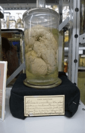
Broca’s brains were again reanalyzed using high resolution magnetic resonance imaging (MRI) by Dronkers et al. to determine the precise location of the lesions as well as other possible areas of damage.8 The results of this novel analysis showed both patients’ lesions were significantly more extensive than originally reported by Broca, providing evidence for inconsistencies between the area originally described by Broca and what is now called Broca’s area. The conclusions of this study have led to significant ramifications: for lesion and functional neuroimaging studies of the brain area historically identified as the major speech center;9 as part of an on-going debate about Broca’s area; and for further exploration of the location of language “centers” in the brain.10 Because of preservation efforts, these brains still exist for future researchers to analyze (see Figures 1 and 2).
The ability to reanalyze the brains studied by Broca demonstrates the need to save the history of neuroscience for future generations. Other examples include the reanalysis of original data sets of the cases of Phineas Gage and Henry Molaison by using modern mapping techniques to compare their lesions to tractography of healthy controls.11 The use of histology demonstration slides from historically important collections, such as those of Sir Charles Sherrington and Sir Wilfrid Le Gros Clark, further demonstrate the importance of saving the history of neuroscience for modern teaching.12
We are facing a dilemma in science because the history of all areas of science, and especially neuroscience, is vanishing. We must develop ways to save it.13 This includes the books, letters, memoirs, and equipment used by scientists and supporting the museums, libraries, and archives that preserve the history of neuroscience. It is also important to teach the value of history for understanding current and future scientific research. All new research is exciting now, but tomorrow it will be history, and the research of tomorrow depends on a broad base of historical findings.14 The development of internet archives helps to make this preservation possible,13 but the real objects must be saved, not just their electronic fingerprints. The history of science is important not just for scientists, but for the cultural and intellectual record that cannot be replaced.
References
- Emiliano Rodríguez Mega, “Second Brazilian Museum Fire In Two Years Reignites Calls For Reform,” Nature 583, (July 2020): 175-176.
- Nora McGreevy, “Why the Cape Town Fire Is a Devastating Loss for South African Cultural Heritage,” Smithsonian Magazine, (April 2021).
- Richard Ovendon, Burning the Books: A History of the Deliberate Destruction of Knowledge (London: John Murray Press, 2020).
- Alison Abbott, “Lost Galileo letter reveals he tried to dodge Inquisition,” Nature 561 (September 2018): 441-442.
- Paul Broca, “Perte de la parole, ramollissement chronique et destruction partielle du lobe antérieur gauche du cerveau,” Bulletin de la Société d’anthropologie de Paris, t. II, (séance du 18 avril 1861): 235-238.
- Paul Broca, “Nouvelle observation d’aphémie produite par une lésion de la moitié postérieure des deuxième et troisième circonvolutions frontales,” Bulletins de la Société Anatomique de Paris 36, (1861): 398-407.
- Jean-Louis Signoret et al., “Rediscovery of Leborgne’s brain: anatomical description with CT scan,” Brain and Language 22, no. 2 (July 1984): 303-319.
- Nina F. Dronkers, Odile Plaisant, M. T. Iba-Zizen and Emmanuel Alain Cabanis, “Paul Broca’s historic cases: High resolution MR imaging of the brains of Leborgne and Lelong,” Brain 130, (May 2007): 1432-1441.
- Adeen Flinker et al., “Redefining the role of Broca’s area in speech,” Proceedings of the National Academy of Sciences of the United States of America 112, no. 9 (February 2015): 2871-5. doi: 10.1073/pnas.1414491112.
- Andrea Gajardo-Vidal et al., “Damage to Broca’s area does not contribute to long-term speech production outcome after stroke,” Brain 144, no. 3 (March 2021): 817-832. doi: 10.1093/brain/awaa460.
- Michel Thiebaut de Schotten et al., “From Phineas Gage and Monsieur Leborgne to H.M.: Revisiting disconnection syndromes,” Cerebral Cortex, 25 no. 12 (December 2015): 4812-4827.
- Bernard S. Chang and Zoltán Molnár, “Practical neuroanatomy teaching in the 21st century,” Annals of Neurology, 77 no. 6 (June 2015): 911-916.
- Lorenzo Lorusso et al., “Neuroscience without borders: Preserving the history of Neuroscience,” European Journal of Neuroscience 48, no. 5 (September 2018): 2099–2109. doi: 10.1111/ejn.14101.
- Richard E. Brown, “Why study the history of neuroscience?” Frontiers in Behavioral Neuroscience 13, article 82 (May 2019). doi: 10.3389/fnbeh.2019.00082.
RICHARD BROWN is a professor of psychology and neuroscience at Dalhousie University and conducts research on the neurobiology of behavior. His research is on mouse models of human neurodegenerative disorders. He is also interested in the history of neuroscience.
THALIA GARVOCK-DE MONTBRUN is a graduate student in psychology at McGill University. Her research involves investigating the neural circuits which underly motivation and reinforcement learning.