Hektoen International

A Journal of Medical Humanities

Cells of an embryo

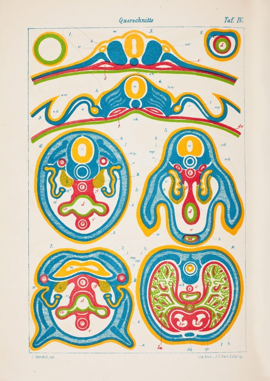
The layers of cells in an embryo, also known as germ layers, develop in stages to create all the parts of the living body. This image from 1874 illustrates exactly that. Showing the differing shapes of differing embryos, but matching the colors of each system across them, creates an effective tool.

For example, the yellow tube near the center of each figure is the neural tube, which will go on to become the central nervous system of the organism. Studying the development of the body in this way can be helpful as well pleasing to the eye.

Colored illustration of the germ layers of an embryo
Illustration from Anthropogenie oder Entwickelungsgeschichte des menschen: gemeinverständliche wissenschaftliche Vorträge über die Grundzüge der menschlichen Keimes- und Stammes-Geschichte by Ernst Haeckel. 1874. Hagstromerbibloteket.

Fall 2019

|

|

Leave a Reply

Your email address will not be published. Required fields are marked *

This site uses Akismet to reduce spam. Learn how your comment data is processed.