 |
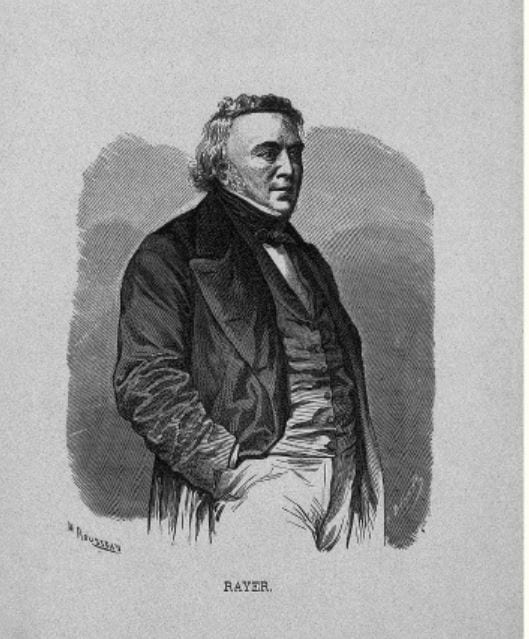
Pierre Rayer occupies a special place in the history of nephrology for his attempt to classify the various diseases that Richard Bright had described in his monumental publication of 1827. With his intern Eugene Napoleon Vigla, he revolutionized the study of kidney diseases by using microscopy to analyze urinary sediments, describing crystals, cells, casts, and yeasts.1,2 Between 1837 and 1841 he published an influential textbook on kidney diseases that was met with international acclaim and has remained a classic ever since.3
At the Hôpital de la Charité, a microscope had become available in 1835 and Rayer promptly set up a program to investigate the urinary findings in the various forms of kidney diseases. His proposed classification was based on clinical findings, urinary microscopy, and gross specimens whenever possible, but not on microscopic examination of sections of the kidney, which at the time had not yet become available. Renal diseases were divided into acute nephritis with many red cells and much albumin in the urine, and chronic albuminuric nephritis corresponding to what is now known as the nephrotic syndrome. There was also mention of suppurative forms of nephritis, with pus cells in the urine, the result of either blood-borne or ascending infection of the kidneys.
Pierre Rayer was born near Caen in the town of Saint Sylvain. It has a street named in his honor but no trace of his family.4 He studied medicine in Paris and in 1812, while still a student, went with five other colleagues to Dijon to help care for Spanish prisoners suffering from typhus.4 He became a hospital intern in 1813 and in 1818 obtained his doctorate with a thesis on the history of pathological anatomy. As a young man, he was denied promotion in academia as punishment for marrying a Protestant, but went on to build a large private practice for mainly wealthy Protestants and Jews.4 In 1823, he was appointed assistant physician at the Academy of Medicine, in 1824 at the Hôpital Saint Antoine, and in 1832 at the Hôpital de la Charité, where he remained until his retirement. In time, he became one of the most prominent and fashionable physicians in Paris, with a wealthy private clientele composed of the most powerful in society.4
His scientific contributions encompassed a remarkably wide range of subjects. He wrote on delirium tremens (1819), sweating or military disease (1822), yellow fever (1822), epidemic nephritis (1839), glanders (1837), and anthrax (1850). His important work on diseases of the skin was published in three volumes in 1826, accompanied by an atlas with 400 illustrations. He is eponymously remembered for describing xanthomas (yellowish nodules on the skin) and Rayer’s disease (an ill-defined chronic disease of jaundice, splenomegaly, and hepatomegaly).
During his long career he received many honors. He was physician to King Louis Philippe (1830-1848) and to Napoleon III, president or member of many learned societies, dean of the school of medicine in 1864, and received the Legion of Honor in that year. A cultured man of wide interests, he is recorded to have been friends with George Sand and other prominent literary figures. He is remembered as “a good, gentle, affable and dignified man” who in the opinion of one of his biographers has “had the misfortune to have been born and to have lived through tempestuous times, when intrigue and violent uprisings were happy bed-fellows.”4
References
- Fogazzi GB and Cameron JS. Urinary microscopy from the 17th century to the present day. Kidney International, 1996, 50: 1058.
- Fogazzi GB and Cameron JS. The introduction of urine microscopy in the clinical practice. Nephrol Dial Transplant, 1995, 9: 410.
- Gabriel Richet. From Bright’s disease to model nephrology: Pierre Rayer’s innovative method of clinical investigation. Kidney International, 1991, 39:787.
- Raymond Molinéry, La vie et l’oeuvre de Rayer, Paris, Médical, 1932, pp. 439–40.
GEORGE DUNEA, MD, Editor-in-Chief
Summer 2018 | Sections | Nephrology & Hypertension

Leave a Reply