
Meena Malhotra
Chicago, Illinois, USA
Redefining the Medical Artist is an exhibition of work by the students, faculty, and staff of the University of Illinois at Chicago (UIC) Biomedical Visualization program. It was held at the International Museum of Surgical Science in Chicago from August 7th to October 16th, 2009. The works featured in this show hope not only to increase public and professional awareness of the many dimensions of this discipline, but to serve as a platform for networking, sharing research ideas and results, and creating new partnerships within the biomedical and scientific community. By seeing and understanding what the field has to offer, we wish to communicate to the viewer what it is we really do and are capable of achieving for medical progress. In other words, we wish to not only dispel the myth that medical illustrators only draw scientific images for textbooks, but to introduce the public to the scope of our abilities. In the tradition of Vesalius’ drawings, we would like to increase your understanding of the discipline of medical illustration through examples of our work.
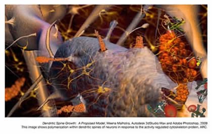
Dendritic Spine Growth
Meena Malhotra
Graduate Student, Biomedical Visualization, UIC
This image is based on the polymerizing of actin molecules into microfilaments that constitute the spines on the dendrites of neurons; this polymerization is one theory on how these spines grow. Given that neurons themselves do not regenerate, some believe that spinal development serves as a basis for memory retention, a process that may be altered by endogenous proteins, such as the ARC protein, or exogenous neurotoxins, such as alcohol. Although the mechanism by which these chemicals may initiate polymerization remains unknown, the image shows a proposed model for ARC-induced spinal growth, increasing the volume of the spine and enhancing the functions relevant to the specific area of the brain.
Visit her website at: http://www2.uic.edu/~mmalho2/
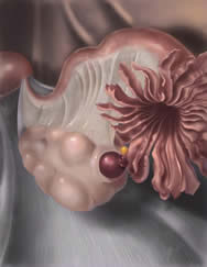
Ovulation
Caroline J. Kaplan
Graduate Student, Biomedical Visualization, UIC
Ovulation was created as part of the Illustrative Techniques course within the BVIS at UIC under course director John Daugherty, MS CMI. This illustration was made to depict a physiological process with special attention to the three aspects of color arrangement: hue, value and saturation. Illustrated in graphite and Adobe Photoshop CS4, an analogous hue scheme with contrasting value and saturation constitute the color arrangement. The image tells a small part of the story behind the female reproductive system, focusing on the moment when a mature egg (ovum) is pushed out of its follicle just prior to rupture during ovulation.
Visit her website at: http://www2.uic.edu/~ckapla4

Surgical Retractors
Nathan McSpadden
Graduate Student, Biomedical Visualization, UIC
This image is a 3D illustration depicting three antiquated retractors from a bygone era in 20th century surgery. Their tight, muscular design suggests they were used for exposing deep structures—perhaps in the abdomen or thigh where many dense layers of tissue are present. Such a wide rake and solid, metal body would be needed to keep these tissues forcefully separated. It is possible that during emergency procedures these retractors made the difference between life and death.
JL Hypospadias Repair
Evelyn Maizels, MD, PhD
Biomedical Visualization, UIC
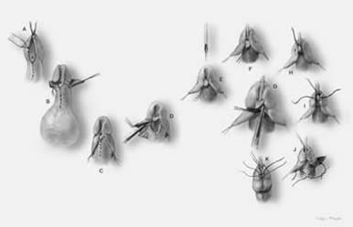
This series was created as part of the Surgical Illustration course within the Biomedical Visualization Program, UIC. Using graphite pencil and Adobe Photoshop, this continuous-tone grayscale digital print is meant to display steps for a novel approach to hypospadias repair, with an intended audience of urological surgeons and residents in training. It can be presented as a figure for inclusion in an article for a Urology journal or as part of the learning module on hypospadias repair within CEVL, the Computer Enhanced Visual Learning Platform.
The steps depicted include:
- A – B: marking the planned skin incisions and raising a skin flap
- E – F: urethral catheterization
- G – J: glans flaps embrace urethra
- K: just prior to completion of procedure
Epstein Barr Virus
Lindsey Brake
Graduate Student, Biomedical Visualization, UIC
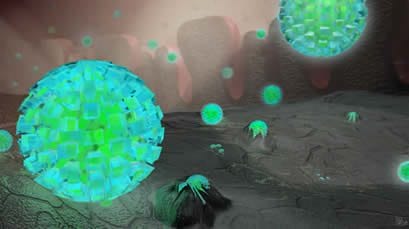
The Epstein-Barr virus is a member of the herpes family, and is one of the most commonly found viruses in humans. Many people become infected during childhood, but the virus remains dormant within the throat and blood cells throughout an individual’s lifetime. There are usually no signs or symptoms when dormant, but, when latent, a person can develop infectious mononucleosis or chronic Epstein-Barr infection. Researchers have also found connections between EBV and Burkitt’s lymphoma, Hodgkin’s disease, and other disorders. EBV easily enters the mouth through contact with an infected person’s saliva, travels through the lingual mucosa, and begins infecting squamous epithelial cells by way of the Lytic Cycle. This piece illustrates the beginning stages of EBV infection, as the virus infiltrates the squamous epithelial cells within the tongue. The cut out slice of the tongue allows the viewer to see the underlying layers of the tongue and how the cells are reacting to the infection. This scene was created in 3d Studio Max and finally composited in Photoshop.
Reach her at: lbrake3@uic.edu.
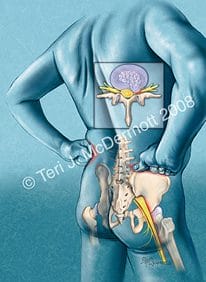
Low Back Pain
Adobe Photoshop CS2
Teri J. McDermott, MA CMI
Alumna and Clinical Assistant Professor, Biomedical Visualization, UIC
Low back pain can be associated with a bulging disc, spinal stenosis, radiculopathy, or another specific spinal cause. This illustration highlights the sciatic nerve and pain caused by a bulging disc.
Commissioned by Jobson Publishing Group
For Cover, U.S. Pharmacist Magazine, May, 2008
Reach her at: Teri@TeriMcDermott.com
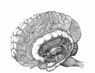
The Limbic System
Traditional pen and ink
LAUREN TIMPSON, Graduate Student, Biomedical Visualization, UIC. This traditional pen and ink illustration shows the structures associated with the limbic system as they lie in the human brain. For clarity, half of the brain and some internal anatomy is not shown. Visit her website at www.timpsonillustrations.com.
MEENA MALHOTRA is a graduate student in the University of Illinois at Chicago’s Biomedical Visualization Program. She is serving as a Guest Curator for the International Museum of Surgical Science’s exhibition Redefining the medical artist. Visit her website at: http://www2.uic.edu/~mmalho2/.
Further reading
Redefining the medical artist
Highlighted in Frontispiece Volume 1, Issue 5 – Fall 2009