
Larry Zaroff
Palo Alto, California, United States
Leonardo Da Vinci (1452–1519)
The surgeon comes to the operating room at seven a.m. for her eight o’clock mitral valve repair. A warm-up. Before any heart operation she always checks the elephants in the room. At that early hour, alone with her elephants, she feels closely connected to them, her better hands. An engineer might differ with the doctor and specify that the elephants are inorganic; a console, a robot with the arms of an octopus that dwells over an operating table, and a monitor, each about six feet tall and three feet wide, a herd. Machines: labeled by the business office, which paid a million plus, as the Da Vinci Surgical System. Both the surgeon and the engineer understand the complex device, know that it is the robot that extends the surgeon’s hands, enabling her to more precisely manipulate instruments through minimal incisions. Of course the engineer and the surgeon identify the name, Leonardo da Vinci, as a painter. They might even recognize his two most famous works, The Mona Lisa and The Last Supper. But if asked why the naming of the elephants, they would not know.*
That Monday morning as she sits at the console with her hands nested in their control compartments to test the moves of each of the robotic arms, duplicating the maneuvers she will use in the heart, she senses someone behind her. Though slightly distracted, she does not turn, assuming one of the nurses. But the voice sounds distant, wave-like, choppy as if his vocal cords were lagging behind his brain: “So this is my namesake—my robot could do much more than translate hands. My robot, in human form, could sit, stand, move its arms and neck, close its mouth, swing his head side-to-side. I’m not impressed, but your optics are satisfactory. You may know that I was the first to describe vision as the result of light entering the eyes.”1,2 When she does turn, she sees nothing, thinks, Weird, I should have had coffee before checking the elephants.
Unhappy, Leonardo, a man of some pride, turned away from the OR thinking, Small stuff, naming a surgical machine because I invented the first robot. What about my work on mechanics and especially my investigations and depictions of human anatomy, far more important.
He could have gone on for hours describing his imaginative inventions from acoustics to military machines, to city planning, to hydrodynamics, to flying, to architecture. During his lifetime, 1452 to 1519, he commented on, enlarged upon, developed virtually every aspect of nature and human activity. His imagination embraced enterprises well beyond his painting. His intellectual curiosity was manic, always going from project to project, often not completing work for which he was paid. In all his ventures, especially the anatomical studies, he saw no separation between art and science, one enhanced the other.
Vesalius, who published his momentous De Humani Corporis Fabrica (The Structure of the Human Body) in 1543, is generally thought of as the father of anatomy; he may not have had that designation if Leonardo had published his drawings in his time.
Leonardo was the first to picture the organs accurately and naturally. Although Renaissance artists studied anatomy, it was the anatomy of the superficial, bony structures and muscles. Leonardo went further, beyond the point when anatomy was useful for his art. Eventually, “Leonardo . . . came to recognize anatomy as more than a subject auxiliary to art.”3 As with most of his investigations, he was driven primarily by the need to learn, to know. He also found that the existing anatomy books were frequently incorrect, “tortuously ponderous . . . and confused” without helpful illustrations to aid the student.4 He did not pursue his anatomical work to benefit doctors, whom he had little use for: “Strive to preserve your health, in which you will be more successful the more you are wary of physicians.”5
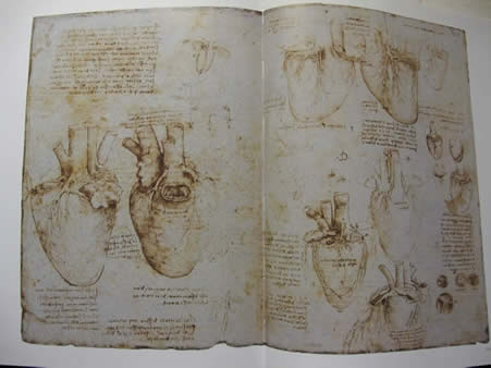
Of his 5,000 found pages of notes and drawings, the majority on mechanics, 190 were devoted to anatomy,** with 50 focused on the heart. These drawings of the heart were accompanied by some 2,000 words, using his mirror handwriting—he was left handed—on the same pages as the pictures.6
In his later years, no longer painting, he focused on the anatomy and physiology of the heart, using the bovine heart as an example. This work, in his last manuscript dated January 9, 1513, is remarkable, original, precise, and elegant, exploring new concepts of function and often disagreeing with Galen.7
In Leonardo’s time, during the Renaissance, Galen’s studies of anatomy remained the norm, the standard, accepted as correct, though his work originated in the second century AD. While Galen argued that the liver, creating blood, was the hub of the vascular system and the heart was merely a churn whose activity heated the body and caused the blood to ebb and flow, Leonardo understood that the heart was central.
Galen thought of the heart as flesh, not a muscle, and alleged, as did all anatomists after him, that the atria were a component of the veins entering the heart. Leonardo pointed out that the heart was a muscle, and as a muscle had its own blood supply and nerves and, for the first time, proved that the atria were distinct chambers whose contractions contributed to the filling of the ventricles.6
Galen, whose dissections were on monkeys, held that the heart dilated by sucking air from lungs, which cooled the heart. The air traveled from the pulmonary veins into the left ventricle where it combined with blood that entered the chamber from the right ventricle through openings in the interventricular septum. However, from experiments in which he inflated lungs, Leonardo proved that air did not enter the pulmonary veins.6
Unaware of the circulation, which Harvey would demonstrate in 1628, Leonardo did not believe blood moved from arteries to veins and accepted Galen’s interventricular openings, though he described them as “imperceptible.”6
Leonardo also disagreed with Galen, showing that the right ventricle pumped blood into the lungs and while the left ventricle did not receive air from the lungs, it propelled blood into the peripheral arteries. But Leonardo did accept Galen’s idea of the lung as an organ to cool blood.8
Leonardo’s contributions to the science of anatomy were both technical and original, exemplified by his studies of the heart. The clarity, detail, and beauty of his drawings were enhanced by his original use of multiple views and cross section visualizations of organs. With precision he described the components and movements of the papillary muscles and chordae tendineae and valves, as well as depicting the coronary arteries and the coronary sinus.8 While Galen hypothesized that dilatation of the heart was more vital than contraction, Leonardo, from his observation of the slaughtering of pigs with a knife inserted into the heart, was able to ascertain that the contraction of the heart in systole produced the pulse. This observation also contrasted with Galen’s belief that arteries dilated independently, suctioning in “vital spirit and producing pulsation.”6
Leonardo rarely acknowledged as facts what was outside his experience. He had to see for himself. Two remarkable studies provide evidence of his powers of observation and his scientific investigations. In a meticulous autopsy of an old man, Leonardo described for the first time atherosclerosis of a coronary artery, which was probably the cause of death. He wrote, “Death . . . to proceed from . . . through lack of blood and deficiency of the artery, which nourishes the heart.”3
Leonardo’s most extraordinary contribution to understanding the circulation was his analysis of how the aortic valve closed, allowing blood flow in only one direction. To study the movements of the valve, Leonardo constructed a glass model of the aortic root. Then using a mix of water and grass seed, he could observe the eddies at the base of the valve in what are now called the sinuses of Valsalva. He believed these eddies initiated closure.8 A similar study in 1969 by Bellhouse verified Leonardo’s experiment as correct.9
The surgeon leans forward, becoming at one with her machine, testing the competence of the repaired mitral valve. No leakage, and the leaflets move easily, blood will not be retarded. The repair is excellent. She smiles and thinks of Da Vinci’s work.
Toby Lester in his Da Vinci’s Ghost describes Leonardo’s drawings and notes as “the most astonishing testament to the powers of human observation and imagination ever set down on paper.”4 I concur and would add that Leonardo’s work on the heart epitomizes his genius.
Notes
* So named by the developer, The Intuitive Surgical Company, reflecting Da Vinci’s early work with robots.
** The number of actual dissections performed by Leonardo is disputed. Most historians count thirty but he surely observed many more autopsies.8
References
- Rosheim, M. (2006). Leonardo’s Lost Robots. New York: Springer.
- Bramley, S. (1992). Leonardo: The Artist and The Man. New York: Penguin Books.
- O’Malley, C. and Saunders, J. (1982). Leonardo Da Vinci on the Human Body. New York: Random House.
- Lester, T. (2012). Da Vinci’s Ghost. New York: Free Press.
- Keele, K. (1952). Leonardo da Vinci on the Movement of the Heart and Blood. Philadelphia: J.B. Lippincott. (Original from pg 287, Anatomical Studies, folio 136r)
- Keele, K. (1951) “Leonardo da Vinci and the Movement of the Heart.” Proceedings of the Royal Society of Medicine, 44, 209–213.
- Clark, K. (1988). Leonardo da Vinci. New York: Viking.
- Clayton, M. and Philo, R. (2012) Leonardo da Vinci Anatomist. Royal Collection Publications
- Boon, B. (2009). “Leonardo da Vinci on atherosclerosis and the function of the sinuses of Valsalva.” Neth Heart J 17(12), 496–499.
LARRY ZAROFF, MD, PhD, has had five careers following his residency and two years in the U.S. Army Surgical Research Unit. He focused for 29 years on cardiac surgery, including a stint as director of the cardiac surgical research laboratory at Harvard. There his work centered on the development of the demand pacemaker. He spent the next 10 years concentrating on climbing and did a first ascent of Chulu West, a 22,000-foot peak on the Nepal-Tibet border. His third life has been at Stanford, where he received a PhD in 2000, and where he teaches courses in medical humanities. His fourth career has been as a writer for the New York Times science section. Until recently, he worked one day a week as a volunteer family doctor. He has received awards as the outstanding faculty advisor for the Human Biology program and in 2006 was honored as Stanford’s Teacher of the Year. He also serves on the editorial board of Hektoen International.
Highlighted in Frontispiece Volume 5, Issue 2 – Spring 2013