
James L. Franklin
Chicago, Illinois, United States

Portions of this paper were read at a meeting of The Chicago Literary Club on March 21, 2016
On October 31, 1794 before a meeting of the Literary and Philosophical Society of Manchester, a young man presented his first scientific paper, “Extraordinary Facts Relating to the Vision of Colours: with observations by Mr. John Dalton.” Dalton (1766–1844)—then 28 years of age—noted, “I was always of (the) opinion, though I might not often mention it, that several colours were injudiciously named.” The paper published in 1798 was the first systematic study of defective color vision. As a Quaker and dissenter from the Anglican Church, Dalton was barred from attending English universities and was largely self-educated. He had recently come to Manchester to teach at the “New College,” a dissenting academy.
Dalton had a condition he referred to as “anomalous vision” which we have come to call color blindness. Sir David Brewster (1781–1868), a Scottish physicist who made many contributions to the field of optics and early photography, is credited with coining the term ‘color-blind.’ As a resident of the United Kingdom, he would have spelled the term “colour-blind.”
“Color blindness” has always been regarded as a troublesome term because it implies a total lack of color vision. Since this is not the case with the common congenital form, “defective color vision” has become the preferred terminology used in the medical field. By the middle of the nineteenth century, to be “color-blind” also came to signify a lack of racial prejudice, but since color vision is our topic, we will not go into this second meaning.
Daltonism
Following the publication of Dalton’s paper in 1798, interest in the subject grew and it became known as “Daltonism.” The term first appeared in print in 1827 by Pierre Prevost of Geneva, a Foreign Member of the Royal Society of London. Well into the twentieth century, the English, French and Spanish retained the term: in France, daltonisme and daltonien, and in Spain, daltonismo.
Even though Dalton remained interested in his defective color vision throughout his life, beyond references to the subject in his correspondence, he never published anything further, and future chemists and Dalton’s admirers objected to having the name of their hero linked to an area that was of minor professional interest to him.
His major scientific contribution was his discovery of the atomic theory of chemistry that is the basis of the Periodic Table.
In his early paper, Dalton recounts how an interest in botany “obliged” him to pay more attention to colors. He often found that when he seriously asked a person if a flower was blue or pink they thought he it was in jest. He continues:
“Notwithstanding this, I was never convinced of a peculiarity in my vision, till I accidentally observed the colour of the flower of the Geranium zonale by candle-light, in the Autumn of 1792. The flower was pink, but it appeared to me almost an exact sky-blue by day; in candle- light, however, it was astonishingly changed, not having then any blue in it, but being what I called red, a colour which forms a striking contrast to blue. ”
While others viewing the flower both in daylight and by candlelight saw no change in color, his brother saw the change in the color of the flower as he did. Dalton enumerates his investigations into the nature of his abnormal vision:
“My observations began with the solar spectrum, or coloured image of the sun, exhibited in a dark room by means of a glass prism. I found that persons in general distinguish six kinds of colour in the solar image; namely, red, orange, yellow, green, blue, and purple. Newton, indeed, divides the purple into indigo and violet; but the difference between him and others is merely nominal. To me it is quite otherwise: —I see only two or at most three distinctions.”
Dalton notes that his was not the first reference to abnormal color vision. A country clergyman named Joseph Huddart in a letter to Joseph Priestly described a shoemaker, Thomas Harris “who could not distinguish colors.” In 1777, Priestly published the letter in the Philosophical Transactions of the Royal Society.
Dalton went on to develop a hypothesis to explain his defective vision:
“It appears therefore almost beyond a doubt, that one of the humours of my eye, and of the eyes of my fellows, is a coloured medium, probably some modification of blue. I suppose it must be the vitreous humour; otherwise I apprehend it might be discovered by inspection, which has not been done. ”
Dalton’s Eyes – Part One
Dalton clung to this explanation of his color defect and instructed his friend and physician Dr. Joseph A Ransome to perform a postmortem examination of his eyes. The day after his death in 1844, Ransome performed the autopsy in the presence of George Wilson, a chemist of Edinburgh and ardent student of defective color vision. Ransome also noted a marked deficiency in the convolutions of Dalton’s brain over the orbital plates. Phrenologists of the time assigned this area to the organ of color.
The eyes were taken to the laboratory of Lyon Playfair, a well-known chemist of Manchester. Ransome collected the fluids of one eye into watch glasses and found them to be “perfectly pellucid.” He shrewdly left the second eye almost intact, slicing off the posterior pole and noted that scarlet and green objects were not distorted in color when seen through the eye. Dalton’s hypothesis was refuted. Ransome dried and stored the eyes. The fragments were preserved along with the Dalton Hall relics in Manchester where they narrowly survived air raids during World War. They were then transferred to the Manchester Museum of Science and Industry.
How the Eye Works
The eye was held in such awe that even before Darwin published The Origin of the Species it was cited as an argument for divine creation. Darwin noted that to suppose the eye “could have been formed by natural selection, seems, I confess, absurd in the highest possible degree.” His task, of course, was to persuade his readers that it was the product of evolution anyway.
The eye gathers information from light that is focused by the cornea and lens on an amazing structure, the retina. The retina transforms light, a physical stimulus, into an electrical signal that is carried to the central nervous system where it is translated into our visual world including the perception of color.
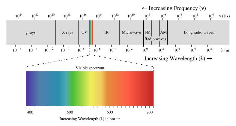
The light we see, “visible light,” is a minute portion of the electromagnetic spectrum, encompassing radio and television waves of great length (hundreds of meters) at one end and X-rays and gamma rays (the diameter of a hydrogen atom) at the other.
Visible light is a narrow band that resides roughly in the middle of the electromagnetic spectrum. [Figure 1] The wavelength of visible light lies between 380 – 760 nm (nm = 10-9 meters). Ultraviolet light resides beyond the short end of the visible spectrum and infrared light beyond the long or red end of the spectrum.[i]
Newton’s Contribution: Primary Colors
In 1666, with the plague raging in Oxford and London, the young Isaac Newton fled to his country home at Woolsthorpe Manor in the county of Lincolnshire. During the next two miraculous years, he developed his theories of optics, gravitation and invented the calculus. At Woolsthrope he performed his experimentum cruces (crucial experiment) on optics. When Newton passed a shaft of daylight through a prism in a darkened room, he identified seven colors: violet, indigo, blue, green, yellow, orange and red – the mnemonic “VIBGYOR” aids in remembering the sequence. The rainbow effect of prisms was already well known, but what he did next was important. He demonstrated that the spectrum of colors could be passed through a second prism and reconstituted to daylight. When he isolated the blue and the red ends of the spectrum and passed them through a second prism they could not be further divided, thus a physical concept of primary colors.
The German mathematician and astronomer Johannes Kepler (1571–1630) in his Harmonice Mundi (The Harmonies of the World) had related planetary orbits to the consonant musical intervals. Newton attempted to equate the seven colors in the spectrum to the musical pitches in an octave. In this he was led astray; his barocentric (circular) model of color vision was incorrect.
Young and Helmholtz’s Contribution: Trichromatic Color Vision
Let us introduce Dalton’s contemporary, the physician and polymath Thomas Young (1773–1829) whose early mastery of multiple languages allowed him in later life to translate the Egyptian Coptic portion of the Rosetta stone, and he contributed to the understanding of Egyptian hieroglyphics making it possible for Jean-François Champolion to decipher the Rosetta stone.
Young made outstanding contributions to our understanding of light, color and vision. His interests were so broad that he often published anonymously fearing that patients and colleagues would think he neglected his medical practice. His earliest scientific paper, published in 1793 when he was twenty years of age, described how the lens of the eye changes shape through the muscular action of the ciliary body allowing for near vision or accommodation. He challenged Newton’s particulate theory of light demonstrating that light behaved as a wave phenomenon and calculated wavelengths for Newton’s seven spectral colors.
Young laid the groundwork for the trichromatic theory of color vision. In 1801 he postulated that receptors or resonators must exist in the retina for three principal colors—red, green and blue—from which all other colors could be produced. It seemed improbable that separate receptors would exist for every conceivable hue throughout the retina. Addressing the issue of Dalton’s abnormal color vision, Young suggested that there might be an “absence or paralysis of those fibers of the retina which are calculated to perceive red.”
At first glance this insight is astounding not only because history would prove him to be correct but also because knowledge of the anatomy of the eye was limited to gross visual inspection. Microscopic examination of the retina still lay in the future. Further reflection on his achievement must take into the account that artists and color theorists dating back to the Renaissance, including Leonardo da Vinci, recognized that with a limited number of primary colors every conceivable hue could be created.
Thomas Young’s three-receptor theory of color vision lay forgotten for some fifty years before it was resurrected in 1851 by another extraordinary scientist, Prussian born Hermann Ludwig Ferdinand von Helmholtz (1821–1894). Famous for his systematic studies of sound and optics, Helmholtz is credited with inventing the ophthalmoscope used today. In resurrecting Young’s conjecture, his name is linked with that of Young as the “Young-Helmholtz trichromatic theory of color vision.”
Maxwell’s Color Box
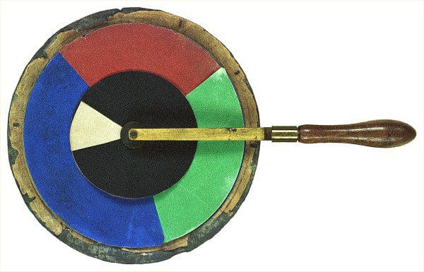
The Scottish physicist James Clerk Maxwell (1831–1879) in the short time allotted him—he died at age forty-eight—is often ranked along with Sir Isaac Newton and Albert Einstein as one of the world’s greatest scientists. Maxwell developed the mathematical concepts allowing for the expression of color in a three-dimensional space. He achieved this using an ingenious top that allowed observers to quantitatively match colors. [Figure 2] He also built a “color box,” a large device of pulleys, mirrors and lenses that objectively tests color matching and the proportion of colors required to achieve a match. These experiments confirmed Young’s hypothesis and Helmholz’s trichromatic theory of color vision. Using red, green and blue, Maxwell was able to match any color. With two colors, a match was not always possible.
Visitors to Maxwell’s London home in the 1860s would meet his wife Katherine, who took a keen interest in her husband’s experiments with the “color box.” Together they collected data on their friends and sought out color-blind people concluding that most of them confused red and green.
Understanding Rods and Cones
The camera was an appealing model for scientists in the nineteenth century for understanding the function of the eye. The retina had to contain some light-sensitive chemical analogous to the silver nitrate film coating the glass slides on which the earliest photographs were captured. German scientists during the second half of the nineteenth century laid the foundations for our understanding of the retina on two levels. They described the cellular elements responsible for vision and identified the light sensitive pigment analogous to the silver nitrate film. The German anatomist and professor at the University of Wüzburg, Heinrich Müller (1820–1864), developed the histologic techniques that allowed him to visualize the previously unseen cellular tissue of the retina. In 1870 his student, Max Schultze (1825–1874) identified the rods and cones as the cellular elements in the retina that were sensitive to light. Through a shrewd deduction in comparative anatomy, Schultze noted that birds that were primarily nocturnal had an abundance of rods while birds that were active during daylight had few rods and many cones. He drew the correct conclusion that the rods were dedicated to night vision and the cones function in daylight.2
Müller also noted the presence of a reddish pigment sensitive to light in the retinas of frogs and squids. In 1876 a student of Schultz, Franz Christian Boll (1849–1879), demonstrated that the pigment was located in the rods and retained its color in the dark but bleached to colorless when exposed to light. When Bolls died at age thirty of tuberculosis, his work was taken up by Wilhelm Kühne (1837–1900) who also succeeded Helmholz as professor of physiology in Heidelberg in 1871 where he worked on the photochemical basis of vision. Kühne extended the study of the pigment to the human retina identifying the pigment as ‘visual purple.’ It would be left to an American, George Wald (1906–1997), to elucidate the biochemistry of ‘visual purple’ naming it rhodopsin (from Greek, rhódin, for rose, and ópsis for sight). Wald, who was Jewish and the son of Polish immigrants, began his studies in Germany during the 1930s working at several famous laboratories. He was forced to return to the United States as a result of the rise of Nazism going first to the University of Chicago and then to Harvard. In 1967, he shared the Nobel Prize in Medicine and Physiology for discoveries concerning the physiological and chemical processes of vision. His lifetime of achievement in the study of the retina included the relationship of Vitamin A deficiency to night blindness, the biochemistry and sensitivity across the visual spectrum of rhodopsin and the related photo pigments present in the retinal cones.3
Of the estimated 130 million photoreceptor cells in the human retina, 120 million are rods and only 6 million are cones. Over 97% of the retina is dedicated to night vision. The evolutionary implication is consistent with the belief that our mammalian ancestors were nocturnal. The rods are more sensitive to light than the cones such that they respond to light that is a billionth the strength of daylight. In daylight the intensity of the light is such that the rods cease to function, they are said to have ‘bleached out.’ At night, our vision is color-blind because only the rod receptors are sending signals to the brain. Night vision is monochromatic. The rods are maximally sensitive to light at a wavelength of 420 nm, corresponding to the blue violet end of the visual spectrum.
The cone receptors are responsible for daylight vision and our perception of color. There are three types of cone cells in the retina: “red,” “green” and “blue.”4 They are more accurately named for the position corresponding to their point of maximal sensitivity in the visual spectrum: ‘L’ for long, ‘M’ for middle and ‘S’ for short wave lengths. To avoid confusion the red, green, and blue terminology is often retained. The point of maximum sensitivity represents the peak sensitivity of a curve plotting the responsiveness of the each type of receptor cell along the wavelengths of the visual spectrum. [Figure 3] Since the curves for each of the receptors overlap, all three receptors respond to a variable degree depending on the light that is falling on the retina. This is the raw material that our central nervous system and visual cortex integrates to generate our perception of color.
It is estimated that of the six million cones in the human retina 99% are “red” or “green;” the “blue” cones represent the remaining one percent. Cones sensitive to the blue end of the spectrum are distributed throughout the retina except in the very center of our visual field designed for maximal visual acuity. This anatomically distinct area, the fovea (“the pit”), is lacking rods and contains only “red” and “green” cones that are more densely packed than in any other part of the retina.5

George Wald plotted the absorbance of rod pigment (black curve), then later the absorbance of cone pigments (red, green, and blue curves)
Notes
- We are exposed to radiant energy from the sun after it is filtered through the atmosphere to a range of 320 nm to 1,100 nm encompassing visible light but including energy in the ultraviolet region and infrared regions of the spectrum.
- Schultz’s work confirmed the earlier observations made by a Czech anatomist, Jan Purkinje (1787–1869), who as an early riser noticed on his walks that the color of his favorite geraniums changed from dark red in the early morning light to pink, as they were lite by the morning sun. Publishing in 1825, he drew the correct conclusion that the eye contained two types of visual equipment, one for daylight and one for dawn and dusk. This phenomenon, known as the Purkinje effect, may call to mind Dalton’s remark about the Geranium zonale.
- The photoreceptor proteins found in the cone cells of the retina that are the basis of color vision as a group are referred to as photopsins. In the L or red cones, photopsin is called ‘erythrodae’ (Greek erythro – red and labe – seeking), in the M or green cons, ‘chlorolabe’ and S or blue cone, ‘cyanolabe.”
- Locating their point of maximum sensitivity in the visual spectrum, the L-cones correspond to greenish-yellow on the visual spectrum, the M-cones correspond to yellowish-green and the S-cones correspond to violet end of the spectrum.
- For this reason, at night, our peripheral vision, while color blind, is more acute than our central vision. Stargazers can confirm this fact by noting that a dimly lit star will be better seen in their peripheral vision than when they attempt to look at it directly.
JAMES L. FRANKLIN is a gastroenterologist and associate professor emeritus at Rush University Medical Center. He also serves on the editorial board of Hektoen International and as the president of Hektoen’s Society of Medical History & Humanities.