
Ceres Alhelí Otero Peniche
Mexico City, Mexico
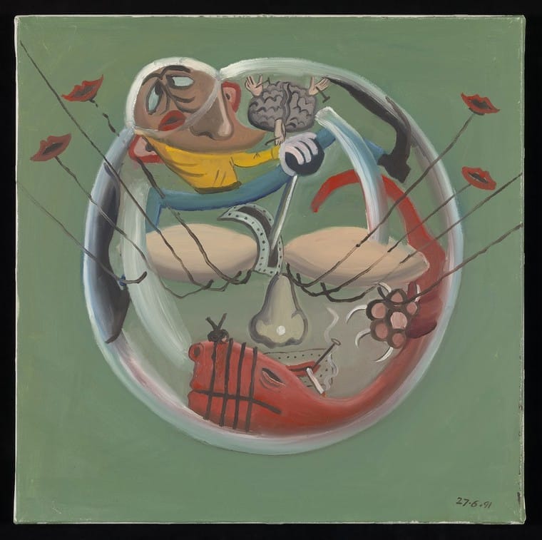
Today I awoke feeling hopeless, disconnected from my body and from my thoughts. All I could sense was the void that my loss of vision represented. I kept thinking how beautiful it would be to see clearly as I opened my eyes. Then suddenly the room began to distort. The shadows slowly transformed into figures, men and women trying to pull off my legs, and then, out of nowhere, a tiger, vibrant and terrifying, ran through my bedroom. I kept telling myself: this is only a dream. But why was I chosen to endure these hallucinations, which grew more vivid as my world grew darker?
This paradox—seeing more as you see less—is the hallmark of Charles Bonnet Syndrome (CBS). Named after the Swiss naturalist who in 1760 described the vivid visions of his grandfather (who was nearly blind from cataracts), it remains one of the most haunting intersections of ophthalmology and neurology, in which patients with visual impairment experience hallucinations despite not being psychiatrically or psychologically impaired.
To understand why a brain deprived of light creates all these hallucinations, we must look at deafferentation theory. Remember that in a healthy visual system, the retina constantly sends a stream of electrochemical signals to the visual cortex in the occipital lobe. This constant stimulus acts as a regulatory mechanism. When conditions like age-related macular degeneration (AMD), glaucoma, diabetic retinopathy, retinal vein occlusion, or cataracts cut off this stream of information, the visual cortex becomes “starved.” This phenomenon is known as denervation supersensitivity.
The neurons responsible for recognizing objects and faces begin to suffer. Without incoming light, the brain opens its own library of memories and images and projects them into the external world. This is like phantom limb syndrome; just as an amputee feels pain in a missing leg, the visually impaired person “sees” with a missing eye.
The specific nature of the hallucinations depends on which areas of the brain are hyper-excitable. If the fusiform gyrus is spontaneously active, the patient sees complex faces. If the parahippocampal gyrus is affected, the patient may see landscapes or buildings. Beyond ocular pathology, CBS is also linked to neurological disruptions. Occipital lobe strokes or low cerebral perfusion pressure can mimic the effects of peripheral vision loss by damaging the primary visual processing centers. In both cases, the result is a dissociation between the eye’s inability to capture light and the brain’s desperate attempt to interpret images.
One of the most tragic aspects of CBS is that it occurs in psychiatrically or psychologically intact individuals, unlike those with schizophrenia or dementia. Many patients remain silent, fearing a psychiatric diagnosis, which only exacerbates isolation. Currently, there is no definitive cure; however, management focuses on treating the underlying cause (such as with cataract surgery) and cognitive behavioral therapy.
Other techniques, such as rapid eye movements (blinking or moving eyes left to right) can sometimes reset the hyper-excited neurons and clear the visual field. Charles Bonnet Syndrome is an evidence of the brain’s incredible plasticity, but is also a reminder that “seeing” is not just a biological function of the eyes, but a complex construction of the mind.
References
- Porter, Daniel. “What is Charles Bonnet Syndrome?” American Academy of Ophthalmology, September 21, 2022. Accessed December 5, 2025. https://www.aao.org/eye-health/diseases/what-is-charles-bonnet-syndrome
- Luis C. Rojas & Bharat Gurnani. “Charles Bonnet Syndrome?” National Library of Medicine, December 6, 2022. Accessed December 5, 2025. https://www.ncbi.nlm.nih.gov/books/NBK585133/
- Forte G, Assaf N, Forte P, Jolly JK. Charles Bonnet Syndrome associated with unilateral vision loss: A new diagnostic perspective. Ophthalmic Physiol Opt. 2025;45(3):681-688. doi:10.1111/opo.13481
- Jones L, Moosajee M. Charles Bonnet syndrome: taking another look at visual hallucinations in sight loss. Ther Adv Ophthalmol. 2025;17:25158414251359588. doi:10.1177/25158414251359588
- Jones L, Jolly JK, Potts J, et al. From research to action: recommendations for Charles Bonnet syndrome care and policy. BMJ Open Ophthalmol. 2025;10(1):e002009. doi:10.1136/bmjophth-2024-002009
- Ucan Gunduz G, Yalcinbayir O, Gelisken O. Awareness of Charles Bonnet syndrome among ophthalmologists: a survey study. Int Ophthalmol. 2025;45(1):207. doi:10.1007/s10792-025-03575-6
CERES ALHELÍ OTERO PENICHE is a fourth-year medical school student at Tecnológico de Monterrey in Mexico City. She is interested in studying nephrology, internal medicine, or microbiology for her academic focus. In her spare time, she enjoys writing, reading, painting, and visiting museums.