
Vidhi Naik
Aberdeen, Scotland
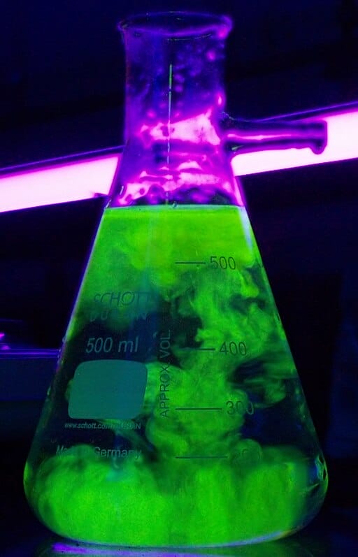
Fluorescein, a strikingly bright orange-yellow liquid, is an essential tool in ophthalmic practice. Its synthesis marked a pivotal moment in the intersection of chemistry and medicine. Created by Adolf von Baeyer, a Nobel prizewinning chemist, in 1871, fluorescein originated as a product of industrial organic chemistry but soon became central to ophthalmic diagnostics. Today, over 150 years after its discovery, it remains a cornerstone in corneal surface assessment and retinal vascular imaging.1,2
Chemical and industrial origins
Following William Henry Perkin’s serendipitous discovery of mauveine, or aniline purple in 1856, chemists explored coal tar derivatives for use as novel colorants.3 German laboratories led by Baeyer, Heinrich Caro, the creator of eosin, and August Kekulé, discoverer of the structure of benzene, dominated this field, investigating reactions of phthalic anhydride with various phenols to produce the phthalein family of dyes.4
Baeyer’s 1871 condensation of phthalic anhydride with resorcinol in the presence of zinc chloride produced a red crystalline compound that emitted brilliant green fluorescence in alkaline solution.1 He named it fluorescein, referencing the mineral fluorite described by Sir George Gabriel Stokes, a contemporary physical scientist in his 1852 paper on fluorescence.4 Sir Stokes detailed this by stating, “I am almost inclined to coin a word, and call the appearance fluorescence, from fluor-spar, as the analogous term opalescence is derived from the name of a mineral.”5 This discovery established fluorescein as the prototype of xanthene dyes, which absorb and emit visible light.
German dye manufacturers such as BASF quickly industrialized fluorescein synthesis.6 The introduction of sodium fluorescein improved water solubility and safety, paving the way for medical use. Bromination of fluorescein yielded eosin, a key histological stain, while related compounds such as rose bengal later expanded the xanthene dye family’s applications in biology and ophthalmology.7
Early medical and ophthalmic applications
In 1882, Swiss ophthalmologist Ernst Pflüger used fluorescein to attempt to understand how the cornea receives cellular nourishment.8 The dye selectively stained damaged tissue, allowing visualization of ulcers, abrasions, and foreign bodies. This simple technique revolutionized corneal diagnostics and remains in daily use worldwide.
With the advent of slit lamp biomicroscopy in the early twentieth century, fluorescein became an essential diagnostic tool. Under cobalt blue illumination, the dye revealed minute surface irregularities. It was soon incorporated into evaluation of the cornea for epithelial defects and tear film assessment, applanation tonometry to evaluate intraocular pressure, and in anterior segment surgery to detect wound leakage.9,10
Fluorescein angiography and retinal imaging
A century after Baeyer’s synthesis, fluorescein again transformed ophthalmology with the introduction of fluorescein fundus angiography (FFA). In 1961, Harold Novotny and David Alvis demonstrated that intravenously injected sodium fluorescein could be imaged as it circulated through the retinal vasculature.11 Using monochromatic excitation and barrier filters, they produced the first dynamic angiograms of the human retina.
Thereafter, FFA enabled visualization of retinal and choroidal circulation in vivo, revolutionizing the diagnoses of diabetic retinopathy, retinal vein occlusion, and choroidal neovascularization.12 Even with the advent of optical coherence tomography (OCT) and OCT angiography, FFA remains the gold standard for detecting vascular leakage and ischemia because of its superior resolution.
Chemical derivatives and biomedical expansion
In the 1940s, fluorescein isothiocyanate (FITC) was synthesized, enabling covalent binding to proteins and antibodies.13 FITC revolutionized cell biology and pathology by allowing fluorescent tagging of biomolecules, and it remains widely used in ophthalmic immunohistochemistry.
Fluorescein’s non-toxicity and detectability also made it valuable in hydrology as a water tracer and in forensic science for detecting biological fluids.14 During World War II, it was even used as a sea rescue marker dye because of its intense visibility.15
Continuing relevance
Fluorescein’s enduring role in ophthalmology exemplifies the bridge between chemistry and clinical innovation. Its dual capacity for surface visualization and vascular imaging has made it indispensable for over a century. Despite technological advances such as OCT, no other agent has matched fluorescein’s diagnostic combination of safety, cost-effectiveness, and dynamic visualization.
Fluorescein’s history mirrors the progress of modern ophthalmology itself. From Baeyer’s laboratory synthesis in 1871 to Hirschberg’s corneal staining and Novotny’s angiography, fluorescein evolved from a chemical curiosity to a universal diagnostic tool. Its story underscores the value of interdisciplinary science, where the pursuit of color in chemistry illuminated the world of vision.
References
- Von Baeyer, Adolf. 1871. “Ueber Eine Neue Klasse von Farbstoffen [on a New Class of Dyes].” Berichte Der Deutschen Chemischen Gesellschaft [Reports of the German Chemical Society] 4 (2): 555–58. https://doi.org/10.1002/cber.18710040209.
- Saine, Patrick J. 1993. “Landmarks in the Historical Development of Fluorescein Angiography.” Journal of Ophthalmic Photography 1 (4): 17–23. https://cdn.ymaws.com/www.opsweb.org/resource/resmgr/history/15-1-04.pdf.
- Plater, M. John, and Andrea Raab. 2017. “Liquid Chromatography-Mass Spectrometry Analysis of Mauveine from the Historical London Suburb of Sudbury (W.H. Perkin’s Home and Factory) and Bradford.” Journal of Chemical Research 41 (8): 441–47. https://doi.org/10.3184/174751917×14967701766969.
- Kekuié, August. 1866. “Untersuchungen Über Aromatische Verbindungen [Investigations on Aromatic Compounds].” Annalen Der Chemie Und Pharmacie [Annals of Chemistry and Pharmacy] 137 (2): 129–96. https://doi.org/10.1002/jlac.18661370202.
- Stokes, George Gabriel. 1852. “On the Change of Refrangibility of Light.” Philosophical Transactions of the Royal Society of London 142 (12): 463–562. https://doi.org/10.1098/rstl.1852.0022.
- Abelshauser, Werner, Wolfgang von Hippel, Jeffrey Allan Johnson, and Raymond G. Stokes. “Becoming a Global Corporation – BASF from 1865 to 1900.” Chapter. In German Industry and Global Enterprise: BASF: The History of a Company, 5–114. Cambridge: Cambridge University Press, 2003.
- Caro, Heinrich. 1892. “Ueber Die Entwickelung Der Theerfarben‐Industrie [on the Development of the Tar Dye Industry].” Berichte Der Deutschen Chemischen Gesellschaft [Reports of the German Chemical Society] 25 (3): 955–1105. https://doi.org/10.1002/cber.18920250399.
- Pflüger, Ernst. 1882. “Zur Ernährung Der Cornea [for the Nutrition of the Cornea].” Klinische Monatsblätter Für Augenheilkunde [Clinical Monthly Journal of Ophthalmology] 20: 70–82. https://babel.hathitrust.org/cgi/pt?id=hvd.hc1enp&seq=83&q1=fluorescein&start=1.
- Cain, W, and R.M. Sinskey. 1981. “Detection of Anterior Chamber Leakage with Seidel’s Test.” Archives of Ophthalmology 99 (11): 2013–13. https://doi.org/10.1001/archopht.1981.03930020889015.
- Keeler. 2024. “Illuminating Insights: A Journey through the History of Fluorescein in Ophthalmology – Keeler Global.” Keeler Global. February 29, 2024. https://www.keelerglobal.com/illuminating-insights-a-journey-through-the-history-of-fluorescein-in-ophthalmology/.
- Novotny, Harold R., and David L. Alvis. 1961. “A Method of Photographing Fluorescence in Circulating Blood in the Human Retina.” Circulation 24 (1): 82–86. https://doi.org/10.1161/01.cir.24.1.82.
- Ruia, Surabhi, and Koushik Tripathy. 2022. “Fluorescein Angiography.” PubMed. Treasure Island (FL): StatPearls Publishing. 2022. https://www.ncbi.nlm.nih.gov/books/NBK576378/.
- Riggs, J.L., R.J. Seiwald, J.H. Burckhalter, C.M. Downs, and T.G. Metcalf. 1958. “Isothiocyanate Compounds as Fluorescent Labeling Agents for Immune Serum.” The American Journal of Pathology 34 (6): 1081. https://pmc.ncbi.nlm.nih.gov/articles/PMC1934794/.
- Smart, Peter L., and S. Laidlaw. 1977. “An Evaluation of Some Fluorescent Dyes for Water Tracing.” Water Resources Research 13 (1): 15–33. https://doi.org/10.1029/wr013i001p00015.
- United States Air Sea Rescue Agency. 1944. “Air Sea Rescue Bulletin V.1 1944-1945: United States. Air Sea Rescue Agency.” Internet Archive. 1944. https://archive.org/details/AirSeaRescueBulletinV1-nsia/page/n5/mode/2up.
DR. VIDHI NAIK is a foundation doctor working in surprisingly sunny northeastern Scotland. She has an interest in ophthalmology, medical history, and advocacy against female genital mutilation (FGM). She aspires to become an ophthalmologist, with a focus on public health.