
Mostafa Elbaba
Doha, Qatar
Water is the essence of life; it serves as the fundamental solvent for many biochemical processes. Different theories have examined how life began on Earth.1 One of the most popular scientific theories suggests that hot chemicals raised through the sea floor enabled a chemical reaction between hydrogen and carbon, producing simple organic compounds. These organic molecules formed more complex organic compounds that became encapsulated in simple cell membranes, which separated the cell as a unit form of life from the surrounding water environment. These complex compounds eventually produced molecules that could carry identity information and DNA.2 These were the first living cells that could grow, divide, and later evolve.3
The phenomenon of osmosis, the movement of a solvent across a semi-permeable membrane from an area of high solvent concentration to an area of low solvent concentration, was first observed in a laboratory setting by Jean-Antoine Nollet (1700–1770). In 1748, Nollet’s experiments demonstrated that water could selectively pass through a membrane, such as a pig’s bladder, while other substances were retained. Osmosis was later described in detail and coined in the early nineteenth century by René Joachim Henri Dutrochet (1776–1847), who observed the phenomenon in both plant and animal tissues while studying the movement of sap.4
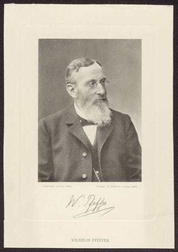
The cell membrane was first conceptualized as a selective barrier that delineated the internal milieu of the cell from its external environment. This barrier was recognized as semipermeable, meaning it allowed the passage of the water as a solvent but restricted the movement of solutes. In the mid-nineteenth century, Moritz Traube (1826–1894) noted that the outer layer must possess semipermeable properties to facilitate the transport of ions, although the precise composition remained unknown.5 In 1877, Wilhelm Pfeffer (1845–1920) proposed the membrane theory of cell physiology. Pfeffer’s work was particularly influential in establishing the importance of this boundary in osmotic phenomena, suggesting that the cell’s surface played a critical role in determining its interaction with the surrounding aqueous environment. The “Pfeffer cell” was an osmometric device he constructed for determining the osmotic pressure of a solution. Pfeffer’s theory posited the existence of an “invisible plasma membrane” surrounding plant cells, which he believed was responsible for their osmotic behavior, drawing parallels between these biological systems and artificial osmometers constructed in the laboratory. In 1889, Ernst Hamburgerconducted experiments on the hemolysis of erythrocytes to determine the permeability of various solutes, further contributing to the understanding of how different substances interact with the cell membrane and affect water movement.6
A crucial step towards understanding the physical nature of the cell membrane was the proposal in 1888 by Georg Hermann Quincke (1834–1924) that the membrane was primarily composed of a thin layer of fat (lipid).7 Quincke’s hypothesis was based on the observation that cells in water tend to form spherical shapes and, when disrupted, break into smaller spheres, a behavior also exhibited by oil. He further noted that thin films of oil could act as semipermeable membranes, aligning with the predicted properties of the cell boundary.8 This idea was further developed by the “lipoid theory of narcosis,” formulated independently by Hans Horst Meyer (1853–1939)9 and Charles Ernest Overton(1865–1933) around the turn of the twentieth century.10 Their research indicated a strong correlation between the lipid solubility of anesthetic molecules and their potency, suggesting that the cell membrane, being lipid in nature, served as a barrier that these molecules had to traverse. Definitive experimental evidence for the lipid bilayer structure emerged in 1924 through the work of Hugo Fricke (1892–1972), who measured the capacitance of erythrocyte solutions, and independently by Evert Gorter (1881–1954) and F. Grendel, who extracted lipids from erythrocytes and determined that the surface area of the extracted lipids, when spread as a monolayer, was twice the estimated surface area of the cells, strongly suggesting a bilayer arrangement.11
The osmosis theory provided a foundational understanding of the cell as a selectively permeable entity, but it lacked the molecular details necessary to explain the high rate at which water was observed to move across certain cell membranes. While the principles of osmosis effectively explained the driving force behind water movement across cell membranes, the physical pathway that allowed water to bypass the membrane, especially at the high rates observed in specific tissues like kidney tubules and red blood cells, remained a key question that the understanding of osmosis alone could not fully address. Moreover, the hydrophilic nature of water and its rapid transport across certain membranes posed a significant challenge to the membrane model, as diffusion through the hydrophobic core of the lipid bilayer was expected to be slow. To address this limitation, several alternative hypotheses were proposed; however, none could fully account for the extremely rapid water transport observed in some specialized cells.12
After establishing the biophysical laboratory at Harvard, Arthur Solomon (1912–2002) and his team conducted pioneering work in the late 1950s on water permeability across red blood cell membranes. They provided compelling evidence that an additional mechanism, beyond simple diffusion, must exist to explain the observed rates of water transport. This body of work strongly hinted at the presence of specific pathways or channels that could facilitate the rapid movement of water across the cell membrane.13

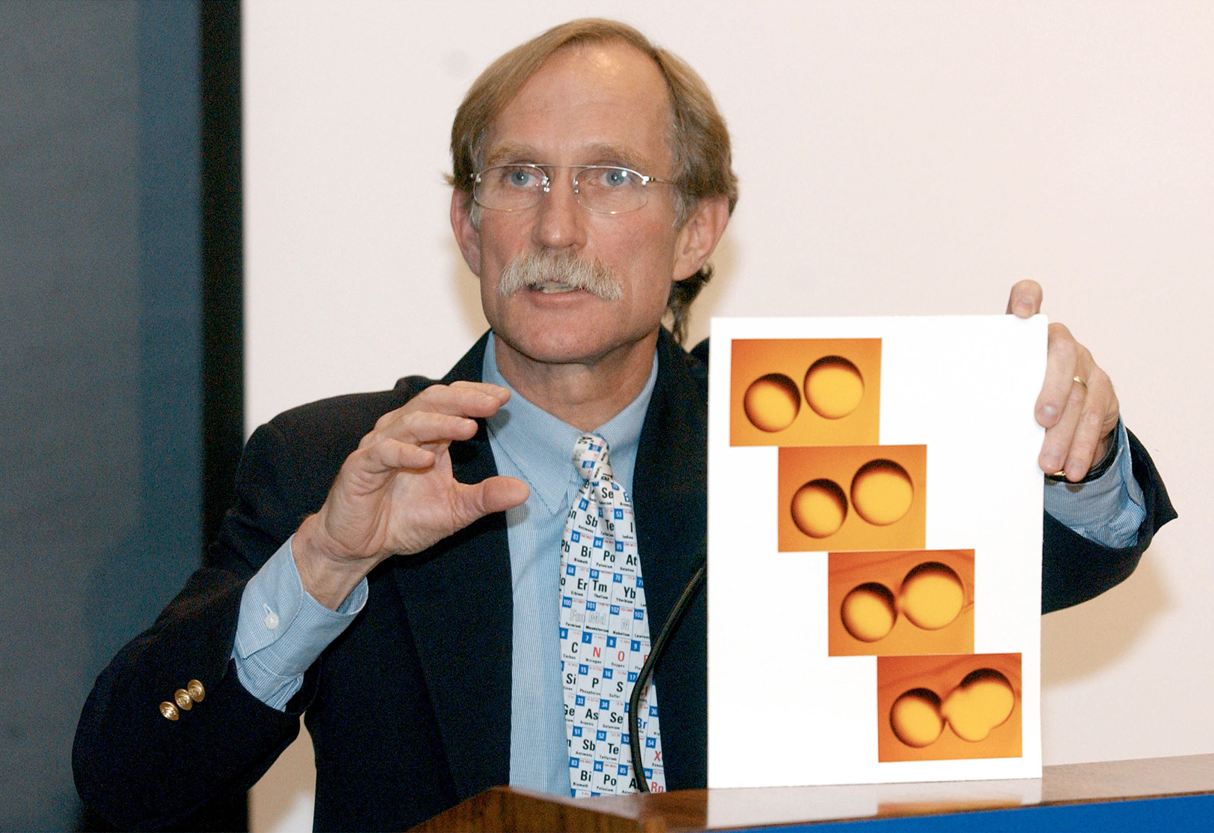
The groundbreaking discovery of water channels—“aquaporins”—by Peter Agre (b. 1949) and his colleagues at Johns Hopkins University in 1992 stands as a remarkable example of good fortune in scientific research. Agre’s primary research focus was on the identification and characterization of the protein responsible for the Rhesus (Rh) blood group antigens in red blood cell membranes.14 During the process of purifying the Rh protein, Agre’s team consistently observed a second protein band on their electrophoretic gels, appearing at a molecular weight of approximately 28 kilodaltons (kDa). Initially considered a potential fragment of the Rh molecule or a mere contaminant, this 28 kDa protein, later designated as Channel-forming Integral membrane Protein of 28 kDa (CHIP28) or simply 28K, persisted in their isolations.15 Intrigued by its abundance, particularly in tissues known for their high water permeability such as red blood cells and kidney tubules, Agre consulted his former hematology professor, John C. Parker, at the University of North Carolina. Parker suggested that this protein might indeed be the long sought-after molecular water channel that had been hypothesized for decades to explain the rapid movement of water across certain cell membranes.16 This insightful suggestion shifted the focus of Agre’s research towards investigating the potential role of this enigmatic 28 kDa protein in water transport.17,18
To rigorously test the hypothesis that CHIP28 functioned as a water channel, Agre collaborated with William Guggino and Gregory Preston at Johns Hopkins University. They employed a well-established assay using frog eggs—Xenopus laevis oocytes. Frog oocytes are known to have a plasma membrane with very low intrinsic water permeability; normally they would not swell significantly or burst when placed in a hypotonic solution like distilled water.19 The researchers injected messenger RNA (mRNA) encoding the CHIP28 protein into the oocytes. The pivotal finding was that after the oocytes expressed the CHIP28 protein and it was transported to their cell membranes, these oocytes exhibited a dramatic increase in water permeability. As shown in the figure; when placed in distilled water, the oocytes expressing CHIP28 rapidly swelled from the influx of water driven by osmosis and eventually burst. In contrast, control oocytes that had not been injected with the CHIP28 mRNA remained intact. This compelling result provided definitive functional evidence that CHIP28 or 28 kDa protein was indeed a protein that mediated rapid water transport across cell membranes, confirming its identity as a water channel.20 This groundbreaking finding was further substantiated by experiments in which the purified CHIP28 protein was reconstituted into artificial lipid vesicles (liposomes), which then showed a significant increase in water permeability compared to liposomes lacking the protein.21
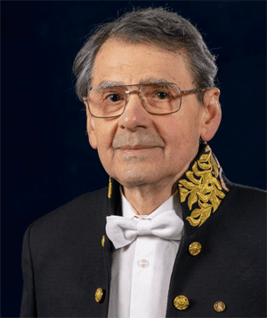
The unknown Rh protein initially known as 28 kDa protein (CHIP28) was later renamed as “aquaporin-1” (AQP1).22 Following the conclusive demonstration of its function, aquaporin became the archetypal member of a newly discovered family of water channel proteins.23 The profound significance of this discovery in unraveling the long-standing mystery of rapid water transport across cell membranes was recognized with the 2003 Nobel Prize in Chemistry,24 which was awarded jointly to Peter Agre and Roderick MacKinnon for work on the structures and mechanisms of membrane ion channels.25 This honor not only acknowledged Agre’s crucial contribution but also highlighted the broad implications of the aquaporin discovery for understanding fundamental biological processes and its potential impact on medicine and biotechnology.26-28
While Peter Agre’s discovery of aquaporin-1 in 1992 is widely celebrated, it is important to acknowledge the earlier work of Gheorghe Benga (born 1944) and his colleagues in Romania.29 In 1986, before Agre’s first publication on the topic, Benga and his coworkers published findings that provided the first evidence of protein-mediated water transport through red blood cell membranes.30 This led to a controversy, with some members of the scientific community arguing that Benga’s pioneering work had not been adequately recognized either by Peter Agre or by the Nobel Prize Committee. Despite this controversy, Agre did acknowledge the contributions of Benga and other pioneers in the field of water transport. In his Nobel lecture, Agre stated that their existence had been suggested by researchers who preceded him by decades.31
References
- Henry, Marc. “Water and the Origin of Life.” Water 16, no. 19 (2024): 2854.
- Seelig, Burckhard, and Irene A. Chen. “Intellectual frameworks to understand complex biochemical systems at the origin of life.” Nature Chemistry 17, no. 1 (2025): 11-19.
- Jordan, Sean F., Hanadi Rammu, Ivan N. Zheludev, Andrew M. Hartley, Amandine Maréchal, and Nick Lane. “Promotion of protocell self-assembly from mixed amphiphiles at the origin of life.” Nature Ecology & Evolution 3, no. 12 (2019): 1705-1714.
- Disalvo, E. Anibal, A. Sebastian Rosa, Jimena P. Cejas, and María de los A. Frias. “Water as a link between membrane and colloidal theories for cells.” Molecules 27, no. 15 (2022): 4994.
- Glater, Julius. “The early history of reverse osmosis membrane development.” Desalination 117, no. 1-3 (1998): 297-309.
- Jacobs, Merkel H. “Early osmotic history of the plasma membrane.” Circulation 26, no. 5 (1962): 1013-1021.
- Keenan, Thomas W. “Historical perspective: milk lipid globules and their surrounding membrane: a brief history and perspectives for future research.” Journal of Mammary Gland Biology and Neoplasia 6 (2001): 365-371.
- De Weer, Paul. “A century of thinking about cell membranes.” Annual Review of Physiology 62 (2000): 919.
- Ling, Gilbert N. “The membrane theory and other views for solute permeability, distribution, and transport in living cells.” Perspectives in Biology and Medicine 9, no. 1 (1965): 87-106.
- Kleinzeller, Arnost. “Charles Ernest Overton’s concept of a cell membrane.” In Current topics in membranes, vol. 48, pp. 1-22. Academic Press, 1999.
- Kalkan, Kübra Tuğçe, and Mukaddes Eşrefoğlu. “The cell membrane: A historical narration.” Cell 8, no. 1 (2020): 81-88.
- Lombard, Jonathan. “Once upon a time the cell membranes: 175 years of cell boundary research.” Biology Direct 9 (2014): 1-35.
- Brown, Dennis. “The discovery of water channels (aquaporins).” Annals of Nutrition and Metabolism 70, no. Suppl. 1 (2017): 37-42.
- Carbrey, Jennifer M., and Peter Agre. “Discovery of the aquaporins and development of the field.” Aquaporins (2009): 3-28.
- Takata, Kuniaki, Toshiyuki Matsuzaki, and Yuki Tajika. “Aquaporins: water channel proteins of the cell membrane.” Progress in Histochemistry and Cytochemistry 39, no. 1 (2004): 1-83
- Dajani, Salah, Anand Saripalli, and Neelam Sharma-Walia. “Water transport proteins–aquaporins (AQPs) in cancer biology.” Oncotarget 9, no. 91 (2018): 36392.
- Finn, Roderick Nigel, and Joan Cerdà. “Evolution and functional diversity of aquaporins.” The Biological Bulletin 229, no. 1 (2015): 6-23.
- Gohar, O. “Aquaporins: The Waterways of Nature.” Modulator 24 (2010): 4-9.
- Agre, P. (2005). Membrane water transport and aquaporins: looking back. Biology of the Cell, 97(6), 355-356.
- Agre, Peter, Landon S. King, Masato Yasui, Wm B. Guggino, Ole Petter Ottersen, Yoshinori Fujiyoshi, Andreas Engel, and Søren Nielsen. “Aquaporin water channels–from atomic structure to clinical medicine.” The Journal of Physiology 542, no. 1 (2002): 3-16.
- Agre, Peter. “The aquaporin water channels.” Proceedings of the American Thoracic Society 3, no. 1 (2006): 5-13.
- Ishibashi, Kenichi, Yoshiyuki Morishita, and Yasuko Tanaka. “The evolutionary aspects of aquaporin family.” Aquaporins (2017): 35-50.
- Madeira, Ana, Teresa F. Moura, and Graça Soveral. “Detecting aquaporin function and regulation.” Frontiers in Chemistry 4 (2016): 3.
- Knepper, Mark A., and Soren Nielsen. “Peter Agre, 2003 Nobel Prize winner in chemistry.” Journal of the American Society of Nephrology 15, no. 4 (2004): 1093-1095.
- The Nobel Prize in Chemistry 2003. NobelPrize.org. Nobel Prize Outreach 2025.
- Verkman, A.S., and Alok K. Mitra. “Structure and function of aquaporin water channels.” American Journal of Physiology-Renal Physiology 278, no. 1 (2000): F13-F28.
- Banerjee, Shohini, Ian M. Smith, Autumn C. Hengen, and Kimberly M. Stroka. “Methods for studying mammalian aquaporin biology.” Biology Methods and Protocols 8, no. 1 (2023): bpad031.
- Vrettou, Charikleia S., Vasileios Issaris, Stelios Kokkoris, Georgios Poupouzas, Chrysi Keskinidou, Nikolaos S. Lotsios, Anastasia Kotanidou, Stylianos E. Orfanos, Ioanna Dimopoulou, and Alice G. Vassiliou. “Exploring Aquaporins in Human Studies: Mechanisms and Therapeutic Potential in Critical Illness.” Life 14, no. 12 (2024): 1688.
- Kuchel, P. W. “The story of the discovery of aquaporins: convergent evolution of ideas—but who got there first.” Cell Mol Biol (Noisy-le-grand) 52, no. 7 (2006): 2-5.
- Benga, Gheorghe. “The first discovered water channel protein, later called aquaporin 1: molecular characteristics, functions and medical implications.” Molecular Aspects of Medicine 33, no. 5-6 (2012): 518-534.
- Figueira, Fernando Faria Andrade, Gustavo Figueira, Raquel Silveira, Viviane Carvalho, Alexandra Seide Cardoso Vidal, Matheus Castro, and Paula Soares. “From water pores to aquaporins: a history of human discoveries… and controversies.” Arquivos de Neuro-Psiquiatria 82, no. S 01 (2024): A058.
MOSTAFA ELBABA is an Egyptian medical doctor, educator, writer and historian. He graduated & granted a master’s degree in pediatrics from Ain Shams University. In 2011, he settled in Hamad Medical Corporation in Qatar. He has a master’s degree in medical education & advanced certification in medical simulation. Apart from medical qualifications, he is a public author & certified in history of medicine, arts, and religions.