
JMS Pearce
Hull, England

A variety of dysraphic states, recorded since antiquity, (Fig 1)1 are caused by the failed closure of the neural tube during the fourth week of embryonic life. They include hydrocephalus, Chiari malformations, and various types of spina bifida with meningocele or meningomyelocele.
Nicolaes Tulp (1593–1674)—subject of Rembrandt’s The Anatomy Lesson—in Observationes Medicae (1641) published six cases and initiated the term spina bifida; his operative attempts failed.
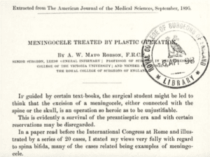
In 1829, Dr. A. Trowbridge of Watertown, New York successfully operated on a boy of eighteen months, otherwise healthy. He observed: “Tumor over lower cervical vertebrae, size of an egg; first the neck was ligated then excised. Wound completely healed and child cured.” He described two similar cervical and sacral “tumours” that were probably meningoceles.2 Other spinal meningoceles treated surgically were reported.3,4 In 1895, the successful, meticulous surgery for a cranial meningocele, the size of a melon, (Fig 2) was described by Mayo-Robson.5 Before his time, aspiration, iodine injections,6 or ligation and excision operations were performed, almost invariably with a fatal outcome.7
A brief excerpt from Mayo-Robson’s paper is of interest: “sutures removed on the sixth day, when the wound was found to be healed, and the patient [illustrated] was discharged cured on the ninth day. When heard of some time afterward it was well and thriving.”
Fully aware of the risks of sepsis and hydrocephalus, he stressed: “unless the operator is prepared to take every pains, both in the details of the operation and in observance of antiseptic precautions, the older rule of non-interference had better be observed.” His operative success was a signal advance in the surgery of simple meningoceles.
 | 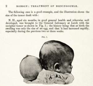 |
| Fig 2. Mayo-Robson, Meningocele. | |
References
- Goodrich, JT. A Historical Review of the Surgical Treatment of Spina Bifida. In: The Spina Bifida. Springer, Milano. 2008. https://doi.org/10.1007/978-88-470-0651-5_1
- Trowbridge A. Boston Medical and Surgical Journal 1829;1 (48), cited by Marcy HO. I. The Surgical Treatment of Spina Bifida. Ann Surg. 1895 Mar;21(3):237-252.
- Roy MC. A Case of Meningocele (Congenital) Complicated with Chronic Hydrocephalus. Ind Med Gaz. 1874 Jul 1;9(7):181-182.
- Westmacott. Spinal meningocele. British Medical Journal 1897; 1579
- Mayo Robson AW. Meningocele treated by plastic operation. Amer J Med Sci 1895;110:280-2.
- Morton JT. Case of spina bifida cured by injection. British Medical Journal 1872;632-3.
- Holmes T. A case of Meningocele in the occipital region. St. George’s Hospital reports 1866;1:39-46.
JMS PEARCE is a retired neurologist and author with a particular interest in the history of medicine and science.
Highlighted Vignette Volume 15, Issue 3 – Summer 2023