Seidumanova Sabrina
Nur-Sultan, Kazakhstan
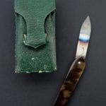
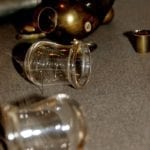
Since time immemorial physicians have treated patients by removing various amounts of blood from the circulatory system. For this purpose they used objects that could cut the skin, such as sharpened pieces of wood, stones, teeth of wild animals, or even the feathers of birds. These tools changed over time, and already by the early twentieth century bloodletting was carried out according to the canons of science of that time. To better see the vein a tourniquet was applied and the vein cut, usually with a lancet, less often with scarificators and bloodletting cupping (Kansas Historical Society, Children’s Illustrated Encyclopedia).
 |
 |
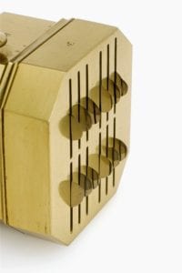 |
| Vaccination lancet, steel and tortoiseshell in leather case, by Mayer and Meltzer of London, 1869–1900 | Bloodletting cups. Josephinum Medical Museum, Vienna, Austria | Brass scarificator with 12 lancets in case, 19th century |
But the most sophisticated way of bloodletting was applying blood-sucking leeches. The procedure was called hirudotherapy. It was facilitated by hirudin, the substance in the leech’s saliva causing more profuse bleeding and making the method less painful by inhibiting the actions of thrombin, one of the blood clotting enzymes (Britannica, 2018). Leeches are sometimes still used today, mainly after microsurgery, to reduce blood congestion in damaged tissue.
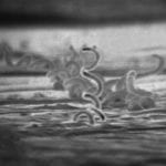
Hirudotherapy had its own complications. The application site could become infected, as described in an 1827 report of a child being infected from a leech previously applied to a patient with syphilis (Quackery). Other patients lost more blood than intended because of hirudin’s systemic blood-thinning properties, such as in the case of a two-year-old girl who died in 1819 from hemorrhage from a single bite of a leech (Quackery). Leeches were not disposable and their repeated use was more dangerous than a single encounter with Bram Stoker’s Dracula!
 |
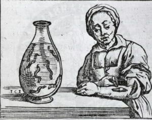 |
| Electron micrograph of Treponema pallidum (syphilis) | A woman standing at a table has placed a leech on her left forearm |
Image Sources
- Vaccination lancet, steel and tortoiseshell, in leather case, by Mayer and Meltzer of London, 1869-1900. Matt black perspex background. Credit: Science Museum, London. CC BY
- Bloodletting Cups. Michelle Enemark, Curious Expeditions on Flickr. 2007. CC BY-NC-SA 2.0. https://www.flickr.com/photos/curiousexpeditions/1772370988/in/photostream/
- Brass scarificator, with 12 lancets, in case, 19th century. Close-up of blades. Three quarter view. White background. Credit: Science Museum, London. CC BY
- Electron micrograph of Treponema pallidum on cultures of cotton-tail rabbit epithelium cells. CDC/ Dr. David Cox. Public Domain. https://phil.cdc.gov/details.aspx?pid=1977
- A woman standing at a table has placed a leech on her left forearm. Illustration in : Bossche, Guillaume van den, Bruxellas, Typis Joannis Mommarti, 1639 Historia medica, in qua libris IV. animalium natura, et eorum medica utilitas esacte & luculenter…. 1639. https://commons.wikimedia.org/wiki/File:Leeching-large.jpeg
Further reading
- “Bloodletting Tools.” Kansas Historical Society, n.d. https://www.kshs.org/kansapedia/bloodletting-tools/10325.
- Children’s Illustrated Encyclopedia. Dorling Kindersley, 2010.
Kang, Lydia, and Nate Pedersen. Quackery: a Brief History of the Worst Ways to Cure Everything. Workman Publishing, 2017. - The Editors of Encyclopaedia Britannica. “Leeching.” Encyclopædia Britannica. Encyclopædia Britannica, inc., November 21, 2018. https://www.britannica.com/science/leeching.
SEIDUMANOVA SABRINA is a student of ISN (International School of Nur-Sultan).
Submitted for the 2019–2020 Blood Writing Contest

Leave a Reply