
JMS Pearce
East Yorks, England
Michelangelo Buonarroti was an exception to the rule that the qualities of many brilliant artists and composers are realized and extolled only after death. He was recognized by contemporaries as a genius, a “Hero of the High Renaissance,” the only artist of whom it was claimed in his lifetime that he surpassed Antiquity.1 Best known among his works are the sculpture of David, his Pietà statues, and the exquisite Sistine Chapel frescoes, including the Last Judgment, created c.1508-1512. It is beyond my ability to adequately appraise Michelangelo’s art; many scholarly works abound.2, 3 How he acquired his anatomical expertise is the focus of this paper.
Born in Caprese in 1475, he considered himself a Florentine, though he lived most of his life in Rome, where he died aged eighty-eight. When thirteen years old he trained first as a painter with Domenico Ghirlandaio, then with the sculptor Bertoldo di Giovannunder. Domenico commended him to Lorenzo de’ Medici, the ruler of Florence.4 At age twenty-one he moved to Rome (1496-1501), where he carved the Vatican Pietà for St. Peter’s Basilica.5 His next moves were to Bologna, then back to Florence, where he sculpted the famous David.
After working in Florence, he was summoned by Pope Julius II to create a sculpted tomb, a project with which he struggled for many years. From 1508 to 1512 he famously painted the vault of the Sistine Chapel with scenes from the Old Testament, from the Creation to the tale of Noah. Immediately celebrated, the Sistine Chapel ceiling displayed many figures in complex, twisting poses, including the fresco of the Last Judgment on the altar wall. Its exuberant use of color became the chief source of the Mannerist style.1
Art and Anatomy
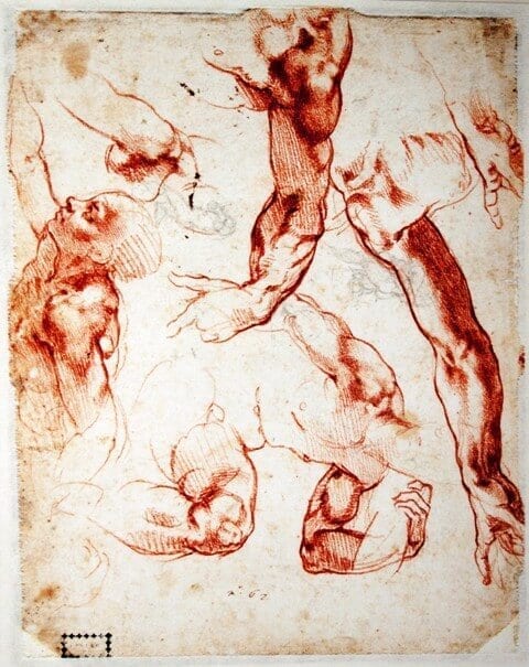
Renaissance artists of the fifteenth and sixteenth centuries, especially those of the Italian schools, studied the human form. The Florentine Academy of Art had an obligatory course in anatomy, in which its students executed drawings from cadavers and skeletons, when available. Few artists performed dissections, but most attended the public dissections of the local physicians and learned from extant anatomical texts. The Church regarded dissection as desecration of the dead, but did intermittently permit dissection of the cadavers of condemned criminals. Many artists of the period tried to study anatomy in detail. In the Lives of Artists, Vasar (1511 – 1574), the famous painter and historian described how important anatomy was to artists:
Again having seen human bodies dissected one knows how the bones lie, and the muscles and sinews, and all order of conditions of anatomy . . . 12
In this respect Michelangelo was not unique. Much has been written in attempts to explain and rationalize the beauty of his works2,3,12 and the way in which they reflected his knowledge of anatomy.6, 7 Michelangelo’s contemporary Leonardo Da Vinci’s (1452-1519) anatomical expertise is comparable. He acquired his first human skull in 1489, and between 1510 and 1511 carried out twenty autopsies at the University of Pavia in collaboration with the professor of anatomy Marcantonio della Torre. By 1513 he had dissected around thirty corpses. Many of Da Vinci’s wonderful drawings and notes were not discovered until the early 1600s; about 600 of his surviving drawings were bound in a single collection and later discovered by William Hunter at Windsor, in the British Royal Collection.
Michelangelo’s anatomy
When aged seventeen, Michelangelo had started his dissections of cadavers from the hospital at the Monastery of Santo Spirito after the death of his mentor Lorenzo de’ Medici. Pope Sixtus IV (r. 1471–1484), himself acquainted with medicine at Bologna,10 permitted dissection in public of condemned criminals, if they were decently buried. However, bodies were also stolen, skinned, and dissected—long before the infamous Burke and Hare, the Edinburgh Body Snatchers of the nineteenth century.
Besides making drawings of dissections, Michelangelo also studied and drew from human models. He became part of the Florentine center of humanism at the Court of Lorenzo de’ Medici.8,9 There he may have met Giovanni Francesco Rustici (1474–1554), a Florentine nobleman, painter, and sculptor taught by Leonardo da Vinci. Michelangelo continually examined dissections and communicated with medical men and their writings. Michelangelo made anatomical studies of the bodies obtained from the Santa Maria del Santo Spirito convent’s hospital. Giorgio Vasari stated:
For the church of Santo Spirito, in Florence, Michael Angelo made a crucifix in wood, which is placed over the lunette of the high altar. This he did to please the Prior, who had given him a room wherein he dissected many dead bodies, zealously studying anatomy.
He probably acquired some of his early knowledge by dissecting with Elia del Medigo, another philosopher physician who was a member of Lorenzo de’ Medici’s circle.10
Michelangelo’s thirst for anatomical knowledge led to selective permission from the Catholic Church to study cadavers. Much has been written in attempts to explain and rationalize the beauty of his works and the way in which they reflected his knowledge of anatomy.7,11 Giorgio Vasari noted that Michelangelo devoted much time to the study of anatomy. In the preface to his Lives, Vasari extols the man and his art:12
And indeed we can affirm with certainty that those do in no way err who call him divine, seeing that he has within his own self embraced the three arts most worthy of praise and most ingenious that are to be found among mortal men,
He was constantly flaying dead bodies, in order to study the secrets of anatomy, thus beginning to give perfection to the great knowledge of design that he afterwards acquired.
. . . In order to be entirely perfect, innumerable times he made anatomical studies, dissecting men’s bodies in order to see the principles of their construction and the concatenation of the bones, muscles, veins, and nerves, the various movements and all the postures of the human body; and not of men only, but also of animals, and particularly of horses, which last he much delighted to keep. (Vasari, Part 11: Summary of Michelangelo’s last years).
By pursuing this discipline of anatomy he acquired great knowledge and skill.13 To quote his pupil Ascanio Condivi:
Through dissection Michelangelo studied every known animal, and did so many human dissections that it outnumbers that of those who are professional in that field. This is a considerable influence that shows in his mastery in anatomy that is not matched by other painters.14
In a letter to Cardinal Ridolfo Pio of Carpi, Michelangelo stated:
It is very certain that the members of architecture depend upon the members of man. Who has not been, or is not a good master of the figure, and especially of anatomy, cannot understand it.
He carried out his own dissections, making molds of muscles in various postures to reveal their surface anatomy, which he applied in painting nude slaves (ignudi), seated near to the sibyls and prophets in panels of the Sistine Ceiling. They can also be seen in the 300 figures in the Last Judgement, which were to represent “the most perfect and well-proportioned composition of the human body in its most varied positions.” (Vasari)
That Michelangelo had an extensive personal knowledge of human anatomy is well established and is obvious in his works.15 In a letter of 1560, in Archivio Buonarroti to Cardinal Ridolfo Pio of Carpi, he remarked: “Who has not been, or is not a good master of the figure, and especially of anatomy, cannot understand it.” Condivi extolled Michelangelo’s anatomical knowledge:
. . . there is no animal whose anatomy he would not dissect, and he worked on so many human anatomies that those who have spent their lives at it and made it their profession hardly know as much as he does.14
So detailed was his knowledge of the human form and movement that later in life he decided to write an anatomical treatise. He sought advice from his close medical friend Realdo Colombo (1516–1559), a surgeon, anatomist, and devotee of Vesalius. Colombo had published in 1559, De re anatomica libri XV. Michelangelo scholars believe that Condivi gave Michelangelo’s ideas of anatomy to Vincenzo Danti, who published the first volume of a proposed huge book on anatomy in 1567 entitled Trattato delle perfette proporzioni. However, Colombo’s text, De Re Anatomica, was printed without Michelangelo’s illustrations, possibly because Michelangelo is known to have destroyed many of his drawings and curiously doubted his own abilities. His plans for the treatise were recorded by Vasari, and by Condivi, who tells us:
. . . He gave up dissection because it turned his stomach so that he could neither eat nor drink with benefit. It is very true that he did not give up until he was so learned and rich in such knowledge that he has often had in mind to write a treatise, as a service to those who want to work in sculpture and painting, on all manner of human movements and appearances and on the bone structure, with a brilliant theory which he arrived at through long experience. He would have done it had he not doubted his powers and whether they were adequate to treat the subject properly and in detail, as someone would who was trained in the sciences and in exposition.14 ( Ch. XI, p.81)
It is claimed that Michelangelo’s anatomical forms demonstrate his use of three sources: firstly, observation of live models and their surface anatomy; secondly, dissection especially of bones and muscles; and thirdly, his reference to the “antique.” There are arguments that his knowledge of dissection is seen in a depiction of deep structures and in surface anatomy. His labeling by names and symbols has also posed problems, but it is clear that both anatomy and its nomenclature were at a primitive stage at the time of his artistic work.
With reference to “The Antique,” Michelangelo is known to have studied inter alia the Belvedere Torso, a copy of an older statue, probably of the early second century BC, housed in the Vatican Museums. In Michelangelo’s The Last Judgement, Saint Bartholomew is shown holding the knife of his martyrdom and his flayed skin. This figure’s torso resembles the Belvedere Torso, which similarly may have inspired several other figures in the Sistine Chapel including the sibyls and prophets, which border the ceiling.
Anatomical curiosities
It has been stated that Michelangelo altered and invented muscles for artistic purposes, but analysis shows that this was rare.16 Contrary to some appraisals, studies of Michelangelo’s art have shown that his portrayals were anatomically remarkably accurate. What appear to be anatomically unusual features are often based on his observations of surface muscles in natural, active postures for purposes of perspective, and what we now call artistic license. Art scholars have suggested that he viewed anatomy—muscles, nerves, and human proportions—as metaphors for certain elements of architecture.17
Unfortunately, he destroyed many of his anatomical sketches and notes. But Suk and Tamargo claim that some of his anatomical illustrations are hidden inside the body of God on the ceiling of the Sistine Chapel, concealed from the eyes of Pope Julius II, and for centuries from countless religious worshipers, historians, and art lovers.18 They conclude that in The Creator separating light from darkness, the creator shows a goiter that has passed unnoticed for almost five hundred years. The neck also resembles the brainstem including the mesencephalon, pons, and medulla.
Earlier, in 1990, Meshberger saw in the fresco Creation of Adam an image that surrounds God and the angels with the shape of a brain.19 The borders in the painting, he asserted, correlate with sulci in the inner and outer surfaces of the brain, the brain stem, the basilar artery, the pituitary gland, and the optic chiasm. It appears that Adam is about to touch God through the contact of their fingertips. Meshberger asserted that Michelangelo was looking for a God representation delivering the intellect to Adam, symbolized by its resemblance to the brain.
In a satirical poem written to a friend, he describes himself as being afflicted with goiter, illustrating the statement with a caricature in the margin of the manuscript.20 It has been said that Michelangelo here portrayed God with a goiter, in his own image.21 Suk and Tamargo interpret the same image as a representation of the brainstem. These differing opinions should prompt caution if not overt skepticism about such imaginative interpretations.
The mantle of the Creator in his painting of the Creator in the Separation of Land and Waters in the Sistine Ceiling is in the shape of the right kidney. Within the mantle are three cherubim watching the massive cosmic movements unfolding where the viewer stands. The shape of the mantle has been described as “a kind of synthesis of the egg and the shell, oval in outline and shell-like in its protective roof.”22 His use of the kidney’s outline suggests to some commentators that Michelangelo was familiar with renal anatomy. Colombo treated him for kidney stones in 1549 and he may have developed gouty arthritis in 1555, making uric acid stones a possibility.10
According to Barreto and Oliveira,23 in the Libyan sibyl painting: “when the vestment which covers the sibyl is inverted, it is possible to notice a huge resemblance between the shoulder articulation with the glenoid cavity (a) and the humeral head (b).” The Creation of Eve is also claimed to show details of the mantle of the Creator, which looks like the shape of the left lung. This deduction and the claimed resemblance are strongly disputed by Kickhofel, who states that they depend only on the power of imagination of those who see the images;24 but Kickhofel’s views too, are rejected by Santos et al.25
Similarly, in Judith and Holofernes, Holofernes has been claimed to resemble the axis vertebra.15
Several authors have tried to identify anatomical shapes such as vertebrae, lungs, joints, brain, and kidney in selected areas of his artwork. Striking as these resemblances may be, the interpretations are somewhat fanciful, and it is hard to imagine why he would have tried to use such devices. These contradictions should make us cautious in interpreting the statements and opinions of the authors.15
Last years
After 1545 Michelangelo devoted himself almost entirely to poetry and architecture. Of many poems in the 1530s and 1540s, approximately 300 survive. In this period he remodeled the Capitol area, the Piazza del Campidoglio, for Pope Paul III. When he died, much of St. Peter’s had been built in its present form. Michelangelo’s marble sculpture after 1545 was limited to two Pietàs. The first, unfinished, was meant for his own tomb. He began the second, the Rondanini Pietà in Milan in 1552, and was working intermittently until February 12, 1564, when overtaken by illness. It carries a transcendental beauty as an expression of love: it contains the figures of Christ and the Virgin, almost merged in a single embrace. He died six days later at his home in Macel de’Corvi, Rome, and was laid to rest at the Basilica di Santa Croce—his chosen place of burial. He had lived to see the publication of the seminal biographies written by Giorgio Vasari12 and Ascanio Condivi.14
Conclusion
The great Michelangelo was primarily responsible for reviving the tradition in post-classical art that sees the human body as the physical manifestation of emotional and spiritual states. Intense feeling requires powerful anatomical forms that Michelangelo acquired from his detailed cadaveric studies and from life.
References
- Michelangelo 1475 – 1564. The National Gallery http://www.nationalgallery.org.uk/artists/michelangelo
- Goldscheider Ludwig. Michelangelo: Paintings, Sculpture, Architecture . London. Phaidon Press 1st edition, 1953
- Bull, George Anthony. Michelangelo: A Biography. New York: Viking, 1995.
- Pettit, Jayne. Michelangelo: Genius of the Renaissance. New York: Franklin Watts, 1998.
- Gilbert, Creighton. Michelangelo. New York: McGraw-Hill, 1967.
- Beck, James H. Three Worlds of Michelangelo. New York: Norton, 1999.
Read more: http://www.notablebiographies.com/Ma-Mo/Michelangelo.html#ixzz3GyoIEFGr - Elkins J. Michelangelo And The Human Form: His Knowledge And Use Of Anatomy. Art History Vol. 7 No. 2 June 1984;176-86.
- Summers, D: Michelangelo and the Language of Art. 1981 Princeton, Princeton University Press,
- Mayor, AH: Artists and Anatomists. 1984 New York, The Metropolitan Museum of Art.
- Eknoyan G. Michelangelo: Art, anatomy, and the kidney. Kidney International 2000; 57: 1190–1201.
- Beck, James H. Three Worlds of Michelangelo. New York: Norton, 1999.
Read more: http://www.notablebiographies.com/Ma-Mo/Michelangelo.html#ixzz3GyoIEFGr - Vasari G. The Lives of the Most Eminent Painters, Sculptors, and Architects. Florence 1550 and 1568 (Michelangelo Buonarroti (1475-1564), translated J.Conaway Bondanaella, P Bondanella) Oxford: OUP 1998:422.
- Holroyd Charles. Michael Angelo Buonarroti, with Translations of the Life of the Master by His Scholar, Ascanio Condivi, and Three Dialogues from the Portuguese by Francisco d’Ollanda. London, Duckworth and Company. P. 16. X111. 1903 http://www.gutenberg.org/files/19332/19332-h/19332-h.html#note_20
- Condivi, A: The Life of Michelangelo. Translation: Baton Rouge, Louisiana State University Press, 1976.
Quotations from Condivi’s Life of Michelangelo are paraphrased based upon the Wohl text and other readings of Michelangelo. - Ellwanger JH, Mohr H, Campos D. Anatomy lessons in the Michelangelo’s works? J. Morphol. Sci., 2012;29 (1):38-43.
- Wallace W. (ed) Michelangelo: Selected Readings. London, Routledge 2013.p.653-4.
- Michelangelo, Anatomy as Architecture, Drawings by the Master. Muscarelle Museum exhibition notes.
http://web.wm.edu/muscarelle/exhibitions/2010/michelangelo.html - Suk I, Tamargo RJ. Concealed neuroanatomy in Michelangelo’s Separation of Light From Darkness in the Sistine Chapel. Neurosurgery, 2010, vol. 66, n. 5, p. 851-861.
- Meshberger FL. An interpretation of Michelangelo’s Creation of Adam based on neuroanatomy. JAMA. 1990 Oct 10;264(14):1837-41.
- Saslow JM. The Poetry of Michelangelo: An Annotated Translation. New Haven: Yale University Press, 1991: 70-2
- Bondeson L, Bondeson A-G. Michelangelo’s divine goiter. J R Soc Med. 2003; 96(12): 609–611.
- De Tolnay, CD. Michelangelo. II. The Sistine Ceiling. 1945 Princeton, Princeton University Press.
- Barreto, G. And Oliveira, Mg. A arte secreta de Michelangelo: uma licão de anatomia na Capela Sistina. São Paulo: ARX, 2004. Cited by Ellwanger , Mohr and Campos.
- Kickhofel, EHP. Uma falsa licão de anatomia ou de um simples caso de impregnacão teórica dos fatos. Scientiae Studia, 2004, vol. 2, n. 3, p. 427-443. Cited by Ellwanger, Mohr, and Campos. (ref 15).
- Santos IP, Rosa JPC, Ellwanger, JH, Molz, P, Rosa, HT, Campos D. Michelangelo’s art on the Sistine Chapel ceiling: sacred representation or anatomy lessons? J. Morphol. Sci., 2013, vol. 30, no. 1, p. 43-48
JMS PEARCE, MD, FRCP, is a retired neurologist and author with a particular interest in the history of science and medicine.
Highlighted in Frontispiece Volume 11, Issue 2– Spring 2019
Leave a Reply